. 2020 Jan 31;19(1):20.
doi: 10.1186/s12943-020-1134-8.

Hsa_circ_001680 affects the proliferation and migration of CRC and mediates its chemoresistance by regulating BMI1 through miR-340

Affiliations

Hsa_circ_001680 affects the proliferation and migration of CRC and mediates its chemoresistance by regulating BMI1 through miR-340

Xiangyu Jian et al. Mol Cancer. .

Erratum in

Abstract

Background: Accumulating evidence indicates that circular RNAs (circRNAs) act as microRNA (miRNA) sponges to directly inhibit specific miRNAs and alter their ability to regulate gene expression at the post-transcriptional level; this mechanism is believed to occur in various cancers. However, the expression level, precise function and mechanism of circ_001680 in colorectal carcinoma (CRC) are largely unknown.

Methods: qRT-PCR was used to detect the expression of circ_001680 and miR-340 in human CRC tissues and their matched normal tissues. Bioinformatics analyses and dual-fluorescence reporter assays were used to evaluate whether circ_001680 could bind to miR-340. Circ_001680 overexpression and knockdown cell lines were constructed to investigate the proliferation and migration abilities in vivo and in vitro through function-based experiments, including CCK8, plate clone formation, transwell, and wounding healing assays. The relationships among circ_001680, miR-340 and BMI1 were investigated by bioinformatics analyses, dual-fluorescence reporter system, FISH, RIP and RNA pull down assays. Sphere forming assays and flow cytometry analyses were used to assess the effect of circ_001680 on the stemness characteristics of CRC cells.

Results: Circ_001680 was more highly expressed in of CRC tissue than in matched adjacent normal tissues from the same patients. Circ_001680 was observed to enhance the proliferation and migration capacity of CRC cells. Furthermore, dual-fluorescence reporter assays confirmed that circ_001680 affects the expression of BMI1 by targeting miR-340. More importantly, we also found that circ_001680 could promote the cancer stem cell (CSC) population in CRC and induce irinotecan therapeutic resistance by regulating the miR-340 target gene BMI1.

Conclusions: Our results demonstrated that circ_001680 is a part of a novel strategy to induce chemotherapy resistance in CRC through BMI1 upregulation. Moreover, circ_001680 may be a promising diagnostic and prognostic marker to determine the success of irinotecan-based chemotherapy.

Keywords: BMI1; Chemotherapy resistance; Has-circ_001680; Irinotecan; Stem cell; miR-340.


Conflict of interest statement

The authors declare that they have no competing interests.

Figures

Fig. 1
Fig. 1
Circ_001680 is overexpressed in colorectal cancer. a Circ_001680 is generated from the B2M gene, located on chromosome 15. b Upper: Sequencing results showing the back-splice junction sequences of circ_001680. Lower: circ_001680 sequences that were PCR-amplified from cDNA and gDNA with divergent primers, respectively. c Relative expression levels of circ_001680 in 42 paired human colorectal cancer tissues. d The Mann-Whitney test was used to detect the relative expression level of circ_001680 in 42 normal and noncancerous tissues(***P < 0.005). e Correlation between circ_001680 expression and the stage of clinical T classification in 42 cases of CRC and noncancerous tissues (**p < 0.01, *p < 0.05). f Graphical illustration of the statistical circ_001680 distribution in CRC patients. (**p < 0.01, *p < 0.05). g qRT-PCR analysis of circ_001680 expression in 11 CRC cell lines
Fig. 2
Fig. 2
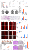
The proliferation and migration of CRC is promoted by circ_001680. a Relative expression of circ_001680 in lentivirus-infected SW480 and HCT116 CRC cell lines. b CCK8 assay comparing the effects of circ_001680 on cell growth between the SW480-vector and SW480-circ_001680 groups (left) and between the SW480-scramble and SW480-circ_001680 inhibitor groups (right). c, d Representative images of the colony formation assay in the indicated cells; d, statistical analysis of the colony formation results. e, f E, Representative transwell results comparing the effects of circ_001680 on cell migration between the SW480-vector and SW480-circ_001680 groups (left) and between the SW480-scramble and SW480-circ_001680 inhibitor groups (right); f, statistical analysis of the transwell assay results. g, h Wound healing assay results showing the differences in migration capacities in the indicated cells at 4 regular intervals (left); statistical analysis of the wound healing assay results (right). i Image of the tumor xenograft model (upper); differences of tumor volume between the SW480-vector and SW480-circ_001680 cells, which were injected into subcutaneous nude mice (lower). j Results of hematoxylin and eosin (H&E) and Ki-67 immunohistochemistry analysis of tumors generated using the tumor xenograft model (left); statistical analysis of Ki-67 positive cells (right). The error bars represent the means±SDs from three independent experiments. **p < 0.01, ***p < 0.005, ****p < 0.001
Fig. 3
Fig. 3
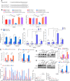
Circ_001680 targets miR-340 and inhibits its transcriptional regulatory activity. a Graphical illustration showing the predicted position of the circ_001680 target on the miR-340 sequence. b Luciferase activity assay showing that circ_001680 influences the luciferase activity of miR-340 3’UTRs in SW480 and HCT116 cells. c qRT-PCR analysis of miR-340 in the indicated lentivirus-infected cells. d qRT-PCR analysis of circ_001680 and miR-340 expression in 43 human colorectal cancer tissues (left); correlation analysis showing that the expression of circ_001680 is negatively correlated with miR-340 (right). e The co-localization between circ_001680 (red) and miR-340 (green) was observed (arrowheads) by fluorescence in situ hybridization in SW480 and HCT116 cells. Cell nuclei were counterstained with DAPI (blue). Scale bar, 5 μm. f According to the RNA pull-down flowchart (left), whole-cell lysates from SW480 and HCT116 cells were incubated with biotinylated probes against circ_001680 or NC; after pull-down, endogenous circ_001680 and miR-340 enrichments were detected by qRT-PCR. Results were presented as the percentage of pull-down to input. g According to the flowchart outlining the experimental procedures (left), The complex containing circ_001680 and miR-340 in indicated cells was immunoprecipitated by anti-Ago2 using RIP (RNA immunoprecipitation) assay and followed by qRT-PCR analyses to detect circ_001680 and miR-340 enrichment (right). h, i Overexpression of miR-340 inhibits the cell growth and migration capacities of SW480 cells induced by circ_001680, as determined by a colony formation assay (h) and a transwell assay (i) in SW480 cells. j Image of the subcutaneous tumor mode. SW480-vector, SW480-circ_001680, SW480-circ_001680 inhibitor and SW480-circ_001680 + miR-340 cells were subcutaneously injected into nude mice separately (n = 6/group). k The tumor volume among the four indicated groups at the indicated time points. l H&E (upper) and Ki-67 immunohistochemistry (IHC, lower) assays were adapted to detect the tumor section. m Statistical analysis of Ki-67-positive cells. All tests were carried out in triplicate. The error bars represent the means ± SDs from three independent experiments. *p < 0.05**, p < 0.01, ***p < 0.005
Fig. 4
Fig. 4
Circ_001680 binds to miR-340 to suppress the expression of BMI1. a Construction of BMI1 3′-UTR WT and mutant (Mut) luciferase reporter vectors. b The dual-luciferase reporter assays indicated that luciferase activity was decreased in CRC cells after cotransfection with the BMI1–3′-UTR-WT and miR-340 mimics. c The whole-cell lysates from SW480 and HCT116 cells were incubated with biotinylated probes against BMI1 or NC; after pull-down, endogenous BMI1 and miR-340 enrichments were detected by qRT-PCR. Results were presented as the percentage of pull-down to input. d The complex containing miR-340 and BMI1 in SW480 and HCT116 cells was immunoprecipitated by by anti-Ago2 using RIP (RNA immunoprecipitation) and followed by qRT-PCR analyses to detect miR-340 and BMI1 enrichment. All tests were carried out in triplicate. e, f qRT-PCR analysis of BMI1 in the indicated cells. g, h Protein expression of BMI1 in the indicated cells as determined by Western blotting. α-tubulin was used as a loading control. The protein expression levels were quantified by comparing the gray level of each band using Quantity One Software. i qRT-PCR analysis of circ_001680, miR-340 and BMI1 expression in the 25 fresh human CRC samples. j Spearman correlation analyses between BMI1 and circ_001680 expression (left), as well as BMI1 and miR-340 (right) expression in 25 fresh human CRC samples (p < 0.01).
Fig. 5
Fig. 5
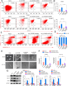
Circ_001680 enhances the colorectal cancer CSC population in vitro. a The number of subpopulations with the CD44+/CD133+ phenotype in the indicated SW480 (upper) and HCT116 (lower) cells. b Statistical results of the number of subpopulation cells with the CD44+/CD133+ phenotype. c The apoptosis assay conducted by flow cytometry in the indicated group. Flow cytometry analyses of SW480 cells treated with 1.0 μM doxorubicin for 24 h. d Statistical analysis of flow results. Annexin-positive/PI-negative cells were analyzed to determine the apoptosis rate. Error bars represent the mean ± SD from three independent experiments. e Typical images from the sphere formation assay among vector, circ_001680, circ_001680 inhibitor and circ_001680 + miR-340. f Graphical illustration showing the sphere formation efficiency from Figure 5C. g Western blot analysis of CD133, SOX2 and CD44 in the indicated lentivirus-infected cells. h qRT-PCR analysis of CD133, CD44 and SOX2 in the indicated cells.
Fig. 6
Fig. 6
Overexpression of circ_001680 suppresses the sensitivity of CRC cells to irinotecan. a The numbers of subpopulation cells with the CD44+/CD133+ phenotype in the indicated SW480 (upper) and HCT116 (lower) cells. b Quantification of cells with the CD44+/CD133+ phenotype is shown in the histogram (*P < 0.05, **P < 0.01). c Typical images from the sphere formation assay in the indicated lentivirus-infected cells treated with or without irinotecan. d Bar graph showing the sphere formation efficiency (*P < 0.05, **P < 0.01). e CCK8 analyzed cell growth capacity in the indicated cells (**P < 0.01). f Apoptosis assay of the indicated cells by flow cytometry (middle). Statistical analysis of the flow cytometry results (right). g Western blot analysis of CD44, CD133, BMI1 and SOX2 in the indicated cells. h qRT-PCR analysis of BMI1, CD44, CD133 and SOX2 in the indicated cells. i Images of the tumor xenograft model (n = 5). j Tumor volume statistics among the four indicated groups on the indicated days. k The tumor weight statistics among the four indicated groups. l H&E and immunohistochemistry analyses of xenograft tumors. IHC staining was performed using antibodies against caspase-3, BMI1, CD44 and SOX-2. The error bars represent the means±SDs from three independent experiments (*P < 0.05, **P < 0.01)

Similar articles

Cited by

References

    1. Brody H. Colorectal cancer. Nature. 2015;521(7551):S1. doi: 10.1038/521S1a. - DOI - PubMed
    1. Siegel RL, Miller KD, Fedewa SA, Ahnen DJ, Meester RGS, Barzi A, et al. Colorectal cancer statistics, 2017. CA Cancer J Clin. 2017;67(3):177–193. doi: 10.3322/caac.21395. - DOI - PubMed
    1. Lieberman DA, Rex DK, Winawer SJ, Giardiello FM, Johnson DA, Levin TR. Guidelines for colonoscopy surveillance after screening and polypectomy: a consensus update by the US multi-society task force on colorectal Cancer. Gastroenterology. 2012;143(3):844–857. doi: 10.1053/j.gastro.2012.06.001. - DOI - PubMed
    1. Sun W, Yang X, Li X, Wang J, Shang R, Li H, et al. Circular RNA: A new star of noncoding RNAs. Cancer Lett. 2015;365(2):141–148. doi: 10.1016/j.canlet.2009.05.004. - DOI - PubMed
    1. Chen I, Chen CY, Chuang TJ. Biogenesis, identification, and function of exonic circular RNAs. Wiley Interdiscip Rev RNA. 2015;6(5):563–579. doi: 10.1002/wrna.1294. - DOI - PMC - PubMed

Publication types

MeSH terms