

Marginal bone resorption of posterior mandible dental implants with different insertion methods
- PMID: 32005142
- PMCID: PMC6995238
- DOI: 10.1186/s12903-020-1019-7
Marginal bone resorption of posterior mandible dental implants with different insertion methods
Abstract
Background: To evaluated the marginal bone loss around dental implants by two insertion methods.
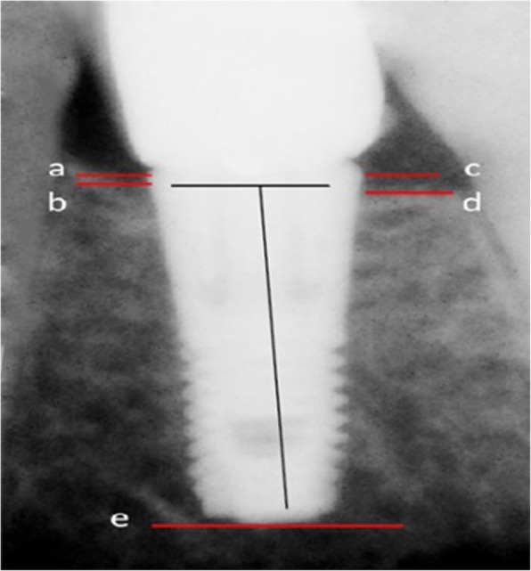
Methods: Eligible patients were divided into two groups; manual and mechanized groups. Peri-apical x-ray using a customized device to standardize the radiographs designed and used to take three periodical radiographs; after surgery, three months, and six months follow up. An independent t-test was used to compare the two groups regarding the average level of marginal bone loss (p < 0.05).
Results: After excluding dropouts, a total of 273 patients (120 males and 153 females, aged between 25 and 67 years old) were included in the study. The average marginal bone loss in the manual insertion method was 0.44 ± 0.84 mm, and 0.59 ± 0.20 mm, and for the mechanized method was 0.51 ± 0.20 mm and 0.67 ± 0.19 mm after three and six months, respectively. There was a significant difference in marginal bone loss after six months between the two groups(p < 0.001). However, no differences were observed after three months (p = 0.24).
Conclusions: Under the condition of this study, both techniques were safe and resulted in an acceptable amount of bone resorption; however, in the manual method, the less marginal bone loss occurred after six months.
Keywords: Bone loss; Dental implant; Implant fixture; Torque devices.
Conflict of interest statement
The authors declare that they have no competing interests.
Figures

Similar articles
-
Does increasing the number of short implants reduce marginal bone loss in the posterior mandible? A prospective study.Br J Oral Maxillofac Surg. 2016 Sep;54(7):731-5. doi: 10.1016/j.bjoms.2016.04.010. Epub 2016 Apr 28. Br J Oral Maxillofac Surg. 2016. PMID: 27131984
-
A prospective multicenter 5-year radiographic evaluation of crestal bone levels over time in 596 dental implants placed in 192 patients.J Periodontol. 2009 May;80(5):725-33. doi: 10.1902/jop.2009.080401. J Periodontol. 2009. PMID: 19405825 Clinical Trial.
-
Ten-year follow-up of dental implants used for immediate loading in the edentulous mandible: A prospective clinical study.Clin Implant Dent Relat Res. 2018 Aug;20(4):515-521. doi: 10.1111/cid.12612. Epub 2018 May 23. Clin Implant Dent Relat Res. 2018. PMID: 29791063
-
Radiologic follow-up of peri-implant bone loss around machine-surfaced and rough-surfaced interforaminal implants in the mandible functionally loaded for 3 to 7 years.Int J Oral Maxillofac Implants. 2004 Mar-Apr;19(2):216-21. Int J Oral Maxillofac Implants. 2004. PMID: 15101592
-
Implant insertion torque and marginal bone loss: A systematic review and meta-analysis.Int J Oral Implantol (Berl). 2020;13(4):345-353. Int J Oral Implantol (Berl). 2020. PMID: 33491366
Cited by
-
The effect of porous compliance bushings in a dental implant on the distribution of occlusal loads.Sci Rep. 2024 Jan 18;14(1):1607. doi: 10.1038/s41598-024-51429-5. Sci Rep. 2024. PMID: 38238380 Free PMC article.
-
Impact of Implant Mesiodistal Distance on Peri-Implant Bone Loss: A Cross-Sectional Retrospective Study.Clin Implant Dent Relat Res. 2025 Feb;27(1):e13442. doi: 10.1111/cid.13442. Clin Implant Dent Relat Res. 2025. PMID: 39844422 Free PMC article.
-
Optimizing the primary stability of dental implants in type IV bone: in-vitro comparison of machine-driven and ratcheting insertion protocols.Eur Oral Res. 2025 Jan 5;59(1):46-52. doi: 10.26650/eor.20241296069. Eur Oral Res. 2025. PMID: 40453411 Free PMC article.
-
Comparison of Vestibular Depth Relapse and Wound Healing After Reconstructive Preprosthetic Surgery Using Cryopreserved Amniotic Membrane and Acellular Dermal Matrix - A Comparative Study.Ann Maxillofac Surg. 2021 Jan-Jun;11(1):12-16. doi: 10.4103/ams.ams_322_20. Epub 2021 Jul 24. Ann Maxillofac Surg. 2021. PMID: 34522647 Free PMC article.
-
Three-Dimensional Evaluation of Alveolar Bone Levels Around Dental Implants and Natural Teeth: A Prospective Study.Cureus. 2024 Oct 9;16(10):e71129. doi: 10.7759/cureus.71129. eCollection 2024 Oct. Cureus. 2024. PMID: 39525190 Free PMC article.
References
-
- Engquist B, Åstrand P, Anzén B, Dahlgren S, Engquist E, Feldmann H, et al. Simplified methods of implant treatment in the edentulous lower jaw: a 3-year follow-up report of a controlled prospective study of one-stage versus two-stage surgery and early loading. Clin Implant Dent Relat Res. 2005;7:95–104. doi: 10.1111/j.1708-8208.2005.tb00052.x. - DOI - PubMed
-
- Lachmann S, Laval JY, Axmann D, Weber H. Influence of implant geometry on primary insertion stability and simulated peri-implant bone loss: an in vitro study using resonance frequency analysis and damping capacity assessment. Int J Oral Maxillofac Implants. 2011;26:347–55. - PubMed
Publication types
MeSH terms
Substances
LinkOut - more resources
Full Text Sources
