. 2020 Jan 31;20(1):8.
doi: 10.1186/s12896-020-0602-y.

Trauma induced tissue survival in vitro with a muscle-biomaterial based osteogenic organoid system: a proof of concept study

Affiliations

Trauma induced tissue survival in vitro with a muscle-biomaterial based osteogenic organoid system: a proof of concept study

Tao He et al. BMC Biotechnol. .

Abstract

Background: The translation from animal research into the clinical environment remains problematic, as animal systems do not adequately replicate the human in vivo environment. Bioreactors have emerged as a good alternative that can reproduce part of the human in vivo processes at an in vitro level. However, in vitro bone formation platforms primarily utilize stem cells only, with tissue based in vitro systems remaining poorly investigated. As such, the present pilot study explored the tissue behavior and cell survival capability within a new in vitro skeletal muscle tissue-based biomaterial organoid bioreactor system to maximize future bone tissue engineering prospects.

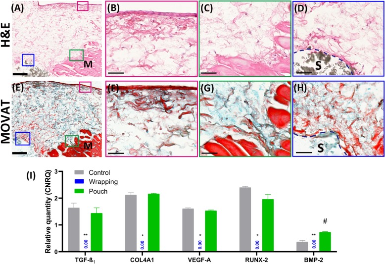
Results: Three dimensional printed β-tricalcium phosphate/hydroxyapatite devices were either wrapped in a sheet of rat muscle tissue or first implanted in a heterotopic muscle pouch that was then excised and cultured in vitro for up to 30 days. Devices wrapped in muscle tissue showed cell death by day 15. Contrarily, devices in muscle pouches showed angiogenic and limited osteogenic gene expression tendencies with consistent TGF-ß1, COL4A1, VEGF-A, RUNX-2, and BMP-2 up-regulation, respectively. Histologically, muscle tissue degradation and fibrin release was seen being absorbed by devices acting possibly as a support for new tissue formation in the bioceramic scaffold that supports progenitor stem cell osteogenic differentiation.

Conclusions: These results therefore demonstrate that the skeletal muscle pouch-based biomaterial culturing system can support tissue survival over a prolonged culture period and represents a novel organoid tissue model that with further adjustments could generate bone tissue for direct clinical transplantations.

Keywords: 3D printed β-TCP/HA; Angiogenesis; Heterotopic implant model; In vitro; Organoid; Pilot study; Tissue survival; Vasculogenesis.


Conflict of interest statement

The authors declare that they have no competing interest.

Figures

Fig. 1
Fig. 1
In vitro wrapping and heterotopic implanted bioceramic pouch model methodology (a-h). The three-dimensional printed macro-porous β-tricalcium phosphate/hydroxyapatite (β-TCP/HA) bioceramic devices (a), for the wrapping or pouch models, were placed in growth medium (DMEM), prior to either wrapping them in rat skeletal rectus abdominis muscle tissue (b-d), or implanting them first in heterotopic extra-skeletal rectus abdominis muscle sites (e-h) of euthanized rats. The implant sites with devices were then harvested, devices embedded in the muscle tissue excised, and subsequently placed in growth medium to be cultured for 5, 15 and 30 days in vitro. (All images within Fig. 1 originate from our own laboratory. Images were not taken from other sources)
Fig. 2
Fig. 2
Comparison of tissue survivability between the two in vitro models in growth medium at day 5, 15 and 30 (a-i). Cells are confined at the interface between muscle and scaffold at day 5 in the wrapping model (a), with a shock silence of tissue-survival related genes (g). Muscle tissue undergo necrosis over time (b) with dying of cells (c, i). In pouch models, initial cell releasing occurs at day 5 (d), leading to successive cell migration and connective tissue formation (e). Vessel structures (f, higher power view) are still present by day 30 in vitro culturing with consistent tissue survival and growth gene expression pattern. Gene expression assays show better tissue survival in the pouch model, especially at day 30 (g-i). Error bars are Mean ± SEM. Ns, non-statistically significant; *, P < 0.05; ***, P < 0.001. H&E staining. M = Skeletal muscle, S = scaffold, CT = connective tissue. Bar: Lower power, 200 μm; higher power, 20 μm
Fig. 3
Fig. 3
Morphology and tissue response to devices in wrapping models and pouch models at day 5 and day 15 (a-h). A considerable amount of fibrils were seen forming into the device (a, f; blue arrows) with some collagen-osteoid formation (green arrow) noticeable at days 5, while the self-adaptation of tissue at the periphery of device was observed in both models (b, e; pink arrows). In contrast, to tissue implanted heterotopically (g, h; blue arrows) the survivability of tissue was compromised in the tissue bag model at days 15, where the muscle tissue on the periphery of the bioceramic device was observed to undergo a type of fragmentation, discontinuing fibrous tissue formation at the interface of the muscle and device (c, d; pink arrows). Movat pentachrome staining was utilized to assess for collagen associated with chondrogenesis and osteogenesis, elastic fibers, muscle and connective tissue. Bars: A, B, D, E, F and H = 100 μm; C and G = 200 μm
Fig. 4
Fig. 4
Microbiological culture results of the 30-day culturing medium with a pouch model. No microbial contamination is detected in the 30-day culturing medium with a pouch model (right plate)
Fig. 5
Fig. 5
Chronological osteogenic-related gene expression pattern in both wrapping and pouch model (a-c). Pouch models showed superior osteogenic differentiation capacity at day 15 (b) and 30 (c) comparing wrapping models. Error bars are Mean ± SEM. Ns, non-statistically significant; **, P < 0.01; ***, P < 0.001
Fig. 6
Fig. 6
Maintenance of vascular structure and potential of angiogenesis in tissue pouch models up to 30 days (a-d). Connective tissue grows into the macropore of the scaffold at the periphery (a, dotted lines show the contour of the macropores), with neurovascular bundle still surviving by 30 days (b). Both transcriptional (c) and translational (d) results suggest the maintenance of angiogenesis capacity with a pouch model by 30 days, whereas the capacity is lost with a wrapping model (P < 0.01 and 0.05, respectively). Error bars are Mean ± SEM. *, P < 0.05; **, P < 0.01. H&E staining. M = Skeletal muscle, S = scaffold, CT = connective tissue, MP = macropores, mp = micropores, BV = blood vessel, N = nerve, C = capillary. Bar: A, 200 μm; B, 50 μm
Fig. 7
Fig. 7
Representative morphology and tissue response to devices in pouch models at day 30 (a-i). Extensive connective tissue forms (a and b) around the scaffold, with comprehensive mucin deposition (e in blue) and fibrils (e in red) evenly distributed in between, consistent with the gene expression pattern showing proliferation and angiogenesis (i). A tissue layer forms at the interface contacting medium (b and f), where fibrous-like cells line at the surface of tissue (b), producing condensed fibers (f in red) underneath. Cells releasing from muscle fiber (c) migrate within the mucin-fibril rich extracellular matrix (g) towards either outer layer or scaffold (d and h). The osteoid (h, area in scarlet) mesh at the interface (dashed lines) between tissue and scaffold indicates the osteogenic transformation of the connective tissue, which is supported by BMP-2 gene expression results (P < 0.05). Error bars are Mean ± SEM. *,#, P < 0.05; **, P < 0.01. H&E staining (a-d); Movat pentachrome staining (e-h). M = Skeletal muscle, S = scaffold. Bar: A and E, 200 μm; B-D, F-H 50 μm

Similar articles

Cited by

References

    1. Amini AR, Laurencin CT, Nukavarapu SP. Differential analysis of peripheral blood- and bone marrow-derived endothelial progenitor cells for enhanced vascularization in bone tissue engineering. J Orthop Res. 2012;30(9):1507–1515. doi: 10.1002/jor.22097. - DOI - PubMed
    1. Anderer U, Libera J. In vitro engineering of human autogenous cartilage. J Bone Miner Res. 2002;17(8):1420–1429. doi: 10.1359/jbmr.2002.17.8.1420. - DOI - PubMed
    1. Denayer T, Stöhr T, Van Roy M. Animal models in translational medicine: validation and prediction. New Horiz Transl Med. 2014;2(1):5–11.
    1. Collier R. Rapidly rising clinical trial costs worry researchers. 2009. - PMC - PubMed
    1. Havers C, Geuder MF. Osteologia nova: sive, Novae quaedam observationes de ossibus, et partibus ad illa pertinentibus: Apud Georgium Wilhelmum Kühnium. 1692.

Publication types

MeSH terms