

Quantitative analysis of facial asymmetry based on three-dimensional photography: a valuable indicator for asymmetrical temporomandibular joint affection in juvenile idiopathic arthritis patients?
- PMID: 32005249
- PMCID: PMC6995089
- DOI: 10.1186/s12969-020-0401-y
Quantitative analysis of facial asymmetry based on three-dimensional photography: a valuable indicator for asymmetrical temporomandibular joint affection in juvenile idiopathic arthritis patients?
Abstract
Background: Juvenile idiopathic arthritis (JIA) can cause osseous deformity in the temporomandibular joint (TMJ) and may impair mandibular growth. This study aimed to evaluate whether facial asymmetry determined clinically or by morphometric analysis of three-dimensional (3D) photographs in JIA patients is associated with an asymmetric affection of theTMJ.
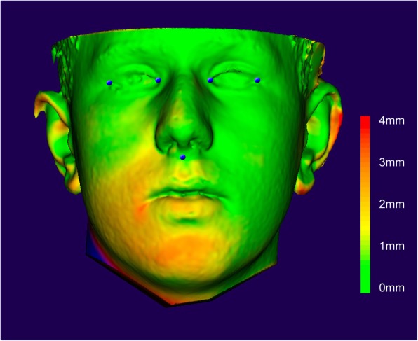
Methods: Of 76 consecutive JIA patients with a mean age of 11.7 years (range: 6.3-17.9), facial asymmetry was evaluated clinically (chin asymmetry, gonion asymmetry), and stereophotogrammetrically with 3D photographs. The facial surfaces were demarcated, then mirrored, superimposed using semi-automated landmarks, and quantitatively assessed (chin asymmetry, Hausdorff distances). Clinical and digital measurements were related to the diagnosis of right and left TMJ involvement derived from magnetic resonance images (MRI).
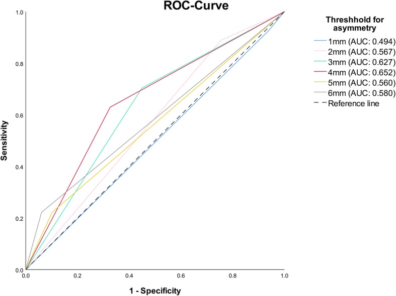
Results: Twenty-seven (34%) patients had an asymmetrical osseous deformity of the TMJ. By clinical evaluation, chin asymmetry was related to asymmetrical osseous destruction (p = 0.02), but gonion asymmetry was not (p = 0.14). In regard to 3D-photograph based morphometric measurements, chin asymmetry was also related to asymmetrical osseous destruction (p = 0.01), but neither the mean (p = 0.06) nor the maximal Hausdorff distance (p = 0.67). Despite the attested significance, none of the chin asymmetry evaluation methods appeared to hold sufficient predictive value (positive predictive values ≤54%; coefficient of determination ≤7%).
Conclusions: For the assessment of facial asymmetry in JIA patients, morphometric measurements originating from 3D-photographs seem to deliver results comparable to the clinical assessment methods. The asymmetry of the face, especially around the chin, appears to be related to asymmetrical TMJ destruction, but none of the investigated measurement methods of the face were able to reliably predict the TMJ affection. Thus, facial asymmetry assessments, both qualitatively in a clinical setting and quantitatively based on 3D-photographs, have limited diagnostic value for TMJ involvement in JIA patients.
Keywords: Facial asymmetry; Juvenile idiopathic arthritis; Morphometric analysis; Stereophotography; Temporomandibular joint; Three-dimensional photography.
Conflict of interest statement
The authors declare that they have no competing interests.
Figures
References
-
- Arvidsson LZ, Fjeld MG, Smith HJ, Flato B, Ogaard B, Larheim TA. Craniofacial growth disturbance is related to temporomandibular joint abnormality in patients with juvenile idiopathic arthritis, but normal facial profile was also found at the 27-year follow-up. Scand J Rheumatol. 2010;39:373–379. doi: 10.3109/03009741003685624. - DOI - PubMed
MeSH terms
LinkOut - more resources
Full Text Sources
Medical
