. 2020 Jan 31;17(1):15.
doi: 10.1186/s12985-020-1290-x.

Design and synthesis of HLA-A*02-restricted Hantaan virus multiple-antigenic peptide for CD8+ T cells

Affiliations

Design and synthesis of HLA-A*02-restricted Hantaan virus multiple-antigenic peptide for CD8+ T cells

Yan Ma et al. Virol J. .

Abstract

Background: Hantaan virus (HTNV) can cause hemorrhagic fever with renal syndrome (HFRS) in humans with severe morbidity and high mortality. Although inactivated HFRS vaccines are given annually for prevention in populations, China still has the highest number of HFRS cases and deaths worldwide. Consequently, vaccination for HFRS requires the development of novel, more effective vaccines. Epitope peptide vaccines have been developed rapidly in recent years and are considered a novel approach for the prevention of infection. Specifically, the multiple antigenic peptide (MAP) design with preferable immunogenicity can arouse a satisfactory immune response for vaccination. However, there are few reports on the design and evaluation of MAP for HTNV.

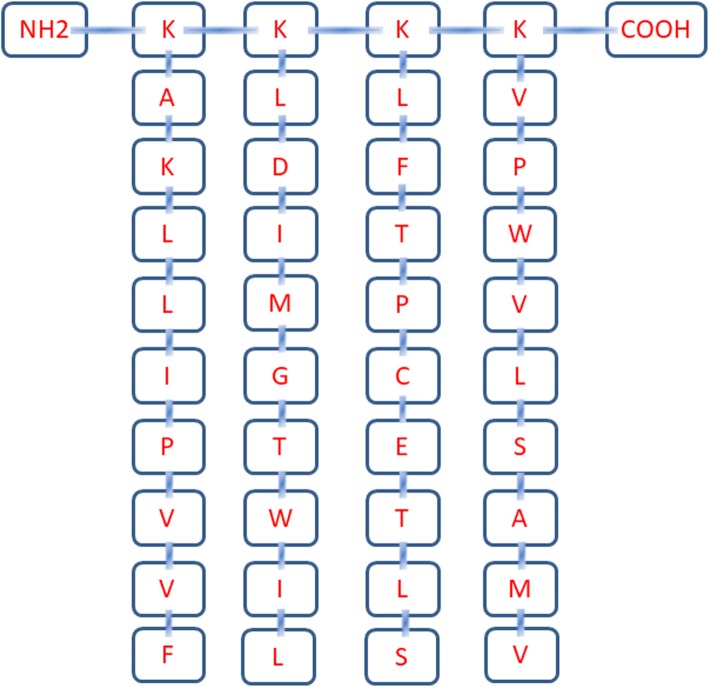
Methods: Three HLA-A*02-restricted 9-mer cytotoxic T lymphocyte (CTL) epitopes on HTNV glycoprotein and one HLA-A*02-restricted 9-mer CTL epitope on the HTNV nucleocapsid, which have been proven to be immunoprotective in our previous study, were selected for the design of HTNV MAP. A four-branched HTNV MAP was evaluated by the IFN-γ-secreting enzyme-linked immunospot assay and proliferation induction capacity of CD8+ T cells and compared with the single HTNV CTL epitope in 17 HLA-A*02+ patients with HFRS. The Mann-Whitney U test was used for comparison of parameters between different subject groups.

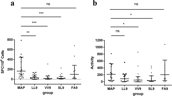
Results: The macromolecular HTNV MAP was designed with a polylysine core and four radially branched single CTL epitope chains. Importantly, HTNV MAP could stimulate CD8+ T cell secretion of IFN-γ in HLA-A*02+ patients with HFRS. The frequency of IFN-γ-secreting CD8+ T cells in the MAP stimulation group was significantly higher than that in the single HTNV CTL epitope stimulation groups (P < 0.005). Meanwhile, the activity of IFN-γ-secreting CD8+ T cells in the HTNV MAP group was also higher than that of the single CTL epitope groups (P < 0.05). Moreover, there was a much stronger ability of HTNV MAP to stimulate CD8+ T cell proliferation compared with that of a single HTNV CTL epitope.

Conclusions: The designed HTNV MAP could induce CTL responses ex vivo and may be considered a candidate for the design and development of novel HTNV peptide vaccines.

Keywords: HLA-A*02; Hantaan virus; Hemorrhagic fever with renal syndrome; Multiple-antigenic peptide; Vaccine.


Conflict of interest statement

The authors declare that they have no competing interests.

Figures

Fig. 1
Fig. 1
The design and synthesis of the HTNV MAP. Four-branched HTNV MAP consists of a polylysine core and attached four HLA-A*02 restricted HTNV 9-mer CTL epitopes. The structure from the amino terminal to the carboxyl terminal is shown. K, lysine; A, alanine; L, leucine; I, isoleucine; P, proline; V, valine; F, phenylalanine; D, aspartic acid; M, methionine; G, glycine; T, tryptophan; C, cysteine; E, glutamic acid; S, serine
Fig. 2
Fig. 2
Comparison of IFN-γ-secreting CD8+ T cell frequency and activity after HTNV MAP and single-peptide stimulation. IFN-γ-secreting cell frequency was detected by enzyme-linked immunospot (ELISPOT) assay. CD4+ T cell-depleted PBMCs from HLA-A*02+ patients with HFRS were stimulated with single peptide (80 μmol/L), MAP (80 μmol/L) or phytohemagglutinin (10 μg/mL). HTNV single CTL epitope LIWTGMIDL, VMASLVWPV, SLTECPTFL, and FVVPILLKA are represented by LL9, VV9, SL9, and FA9, respectively. a The frequency of IFN-γ-secreting CD8+ T cells was detected. All cell frequencies were converted to the number of spot-forming cells per 1 × 106 CD4+ T cell-depleted PBMCs (SFCs/106 cells). b The activity of IFN-γ-secreting CD8+ T cells was measured. The activity was evaluated as the average of the spot size and intensity for each well. Each black dot represents one patient sample. The Mann-Whitney U test was used to determine the difference between two groups. The black line indicates the median and corresponding interquartile range (IQR). *P < 0.05, **P < 0.01, ***P < 0.001, ns indicates no significant difference
Fig. 3
Fig. 3
Representative CFSE staining of CD8+ T cell proliferation stimulated with HTNV MAP and single peptide. Isolated PBMCs from HLA-A*02+ patients with HFRS were labeled with CFSE (5 μM) and stimulated with HTNV MAP or single epitope LL9, VV9, SL9 and FA9. The cells were stained with anti-CD3 PerCP-Cy5.5 and anti-CD8 PE mAb after 5 days of culture. a The scatter plot for the flow cytometry analysis of cell proliferation from one representative HLA-A*02+ HFRS patient after stimulation with different groups of stimuli when gated on CD3+ cells. The upper left quadrant shows that the CFSE fluorescence intensity was reduced in cells representing the proliferation percentages. The number indicates the percentage of cells in the boxed area. b The histogram shows the peak shift of the CFSEdimCD3+CD8+T cells in different stimulation groups. The degree of shift to the left of the curve reflects the proliferation percentage of CD8+ T cells. The SEB stimulation group was used as a positive control, and the no stimulation group was used as a negative control

Similar articles

Cited by

References

    1. Kruger DH, Figueiredo LT, Song JW, Klempa B. Hantaviruses--globally emerging pathogens. J Clin Virol. 2015;64:128–136. doi: 10.1016/j.jcv.2014.08.033. - DOI - PubMed
    1. Vaheri A, Strandin T, Hepojoki J, Sironen T, Henttonen H, Mäkelä S, et al. Uncovering the mysteries of hantavirus infections. Nat Rev Microbiol. 2013;11(8):539–550. doi: 10.1038/nrmicro3066. - DOI - PubMed
    1. Witkowski PT, Perley CC, Brocato RL, Hooper JW, Jürgensen C, Schulzke JD, et al. Gastrointestinal tract as entry route for hantavirus infection. Front Microbiol. 2017;8:1721. doi: 10.3389/fmicb.2017.01721. - DOI - PMC - PubMed
    1. Watson DC, Sargianou M, Papa A, Chra P, Starakis I, Panos G. Epidemiology of hantavirus infections in humans: a comprehensive, global overview. Crit Rev Microbiol. 2014;40(3):261–272. doi: 10.3109/1040841X.2013.783555. - DOI - PubMed
    1. Li Z, Zeng H, Wang Y, Zhang Y, Cheng L, Zhang F, et al. The assessment of Hantaan virus-specific antibody responses after the immunization program for hemorrhagic fever with renal syndrome in Northwest China. Hum Vaccin Immunother. 2017;13(4):802–807. doi: 10.1080/21645515.2016.1253645. - DOI - PMC - PubMed

Publication types

MeSH terms