. 2020 Apr;214(4):941-959.
doi: 10.1534/genetics.120.303085. Epub 2020 Jan 31.

The Role of pkc-3 and Genetic Suppressors in Caenorhabditis elegans Epithelial Cell Junction Formation

Affiliations

The Role of pkc-3 and Genetic Suppressors in Caenorhabditis elegans Epithelial Cell Junction Formation

José G Montoyo-Rosario et al. Genetics. 2020 Apr.

Abstract

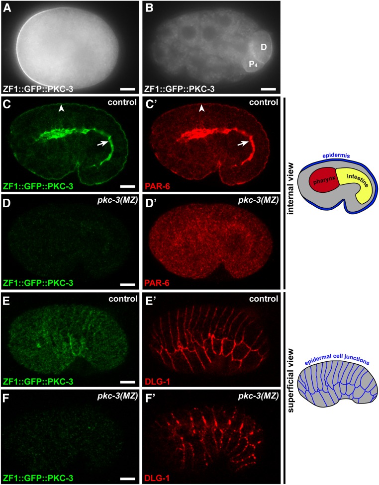
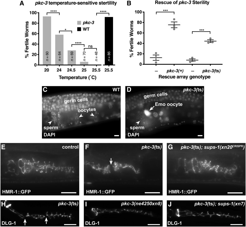
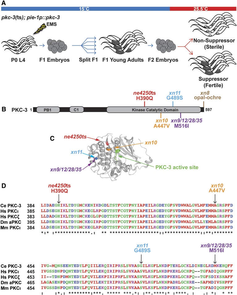
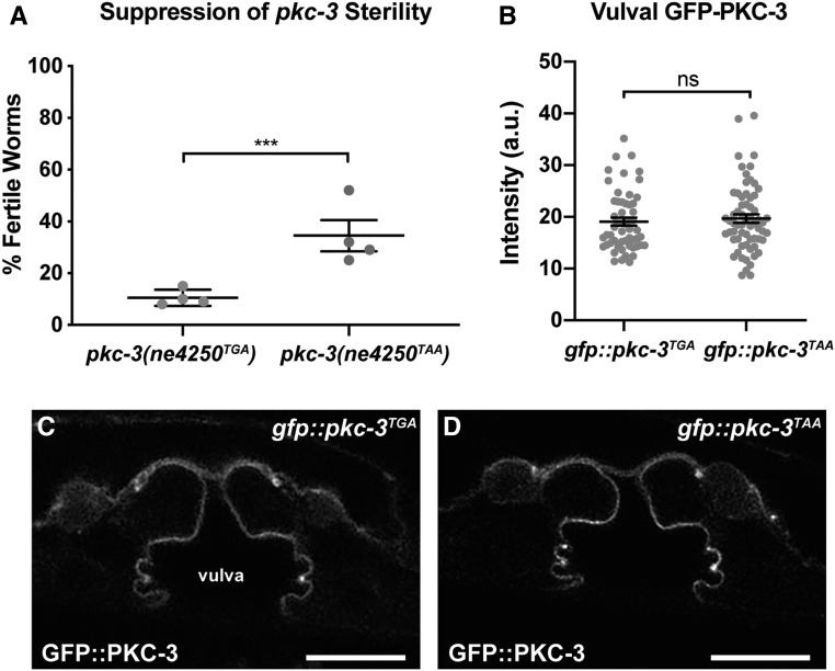
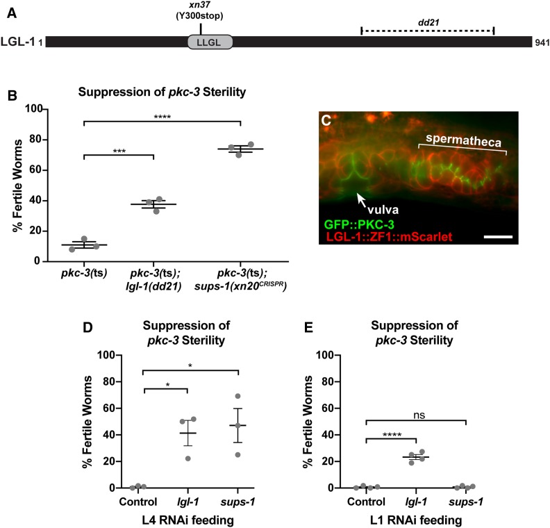
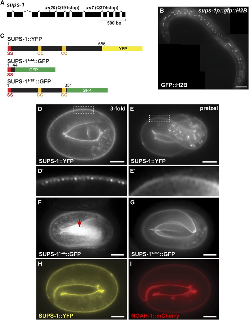
Epithelial cells form intercellular junctions to strengthen cell-cell adhesion and limit diffusion, allowing epithelia to function as dynamic tissues and barriers separating internal and external environments. Junctions form as epithelial cells differentiate; clusters of junction proteins first concentrate apically, then mature into continuous junctional belts that encircle and connect each cell. In mammals and Drosophila, atypical protein kinase C (aPKC) is required for junction maturation, although how it contributes to this process is poorly understood. A role for the Caenorhabditis elegans aPKC homolog PKC-3 in junction formation has not been described previously. Here, we show that PKC-3 is essential for junction maturation as epithelia first differentiate. Using a temperature-sensitive allele of pkc-3 that causes junction breaks in the spermatheca and leads to sterility, we identify intragenic and extragenic suppressors that render pkc-3 mutants fertile. Intragenic suppressors include an unanticipated stop-to-stop mutation in the pkc-3 gene, providing evidence for the importance of stop codon identity in gene activity. One extragenic pkc-3 suppressor is a loss-of-function allele of the lethal(2) giant larvae homolog lgl-1, which antagonizes aPKC within epithelia of Drosophila and mammals, but was not known previously to function in C. elegans epithelia. Finally, two extragenic suppressors are loss-of-function alleles of sups-1-a previously uncharacterized gene. We show that SUPS-1 is an apical extracellular matrix protein expressed in epidermal cells, suggesting that it nonautonomously regulates junction formation in the spermatheca. These findings establish a foundation for dissecting the role of PKC-3 and interacting genes in epithelial junction maturation.

Keywords: aPKC; adherens junction; cell polarity; kinase; stop codon; suppressor.


Figures

Figure 1
Figure 1
PKC-3 is required for PAR-6 localization and epithelial junction maturation. (A) One-cell stage embryo. ZF1::GFP::PKC-3 is enriched at the anterior cortex. (B) 24-cell stage embryo. ZF1::GFP::PKC-3 has degraded from all embryonic cells except for the germline precursor (“P4”) and its recently born sister cell (“D”). (C–C’) ZF1::GFP::PKC-3 and PAR-6 localization in a central plane of a fixed and immunostained comma-stage embryo. Both proteins colocalize at apical surfaces of epithelia, including the epidermis (arrowhead) and intestine (arrow); see schematic at right for positions of major classes of epithelial cells. (D–D’) ZF1::GFP::PKC-3 is not detected in a pkc-3(MZ) embryo and PAR-6 apical enrichment is lost. (E–E’) ZF1::GFP::PKC-3 and DLG-1 in a superficial view of the epidermis in a comma stage embryo. DLG-1 forms continuous belts around each epidermal cell; see schematic at right. (F–F’) ZF1::GFP::PKC-3 and DLG-1 in a pkc-3(MZ) embryo; maternal ZF1::GFP::PKC-3 is degraded and DLG-1 accumulates in apicolateral clusters rather than continuous belts. Bar, 10 µm.
Figure 2
Figure 2
pkc-3(ts) causes sterility and spermathecal junction gaps that are rescued by suppressors. (A) Fertility of pkc-3(ne4250) worms shifted at the L1 stage to the indicated temperature (gray bars), and fertility of wild-type worms at 25.5° (black bar). ****P ≤ 0.001, *P ≤ 0.05, ns, not significant, Fisher’s exact test. (B) Rescue of sterility of pkc-3(ne4250) worms, shifted at the L1 stage to 25.5°, by extrachromosomal arrays expressing pkc-3(+) (xnEx533) or pkc-3(ne4250) (xnEx534) with sur-5::gfp cotransformation marker. Array-negative control worms are the siblings of array-bearing worms. Gray dots indicate mean fertility from one of three individual experiments, horizontal line is the mean of means, and error bars are the SEM. Sample sizes from three experiments: xnEx533 control (n = 98, 113, 87); xnEx533 (n = 101, 105, 110); xnEx534 control (n = 89, 111, 98); xnEx534, (n = 93, 104, 85). ***P ≤ 0.001, unpaired Welch’s t-test. (C and D) DAPI-stained gonads in fixed young adult wild-type and pkc-3(ts) hermaphrodites. Germ cells (mitotic or early meiotic), oocytes, and sperm are indicated. An endomitotic (Emo) oocyte nucleus in pkc-3(ts) is shown in (D, arrow). (E–G) Endogenously tagged HMR-1::GFP in adherens junctions of the spermatheca of live L4 stage worms of the indicated genotype at 25.5°; control worms are wild-type worms expressing HMR-1::GFP. A break in junctional HMR-1::GFP in a pkc-3(ts) worm is indicated (F, arrow). (H–J). Spermathecal junctions in fixed L4 larvae of the indicated genotype stained for DLG-1; all genotypes also contain the pie-1p::pkc-3 (xnIs131) transgene to circumvent maternal-effect lethality. Breaks in the spermathecal junctions of a pkc-3(ts) mutant are indicated (H, arrow). Bar, 10 µm.
Figure 3
Figure 3
Intragenic pkc-3(ts) suppressors are in the kinase domain and stop codon. (A) Design of the pkc-3 suppressor screen. The pie-1p::pkc-3 (xnIs131) transgene expresses pkc-3 only maternally and was used to bypass the requirement for pkc-3 in anterior–posterior polarity in the early embryo. (B) PKC-3 protein depicting conserved domains and locations of the temperature-sensitive mutation and intragenic suppressors. (C) Structure of the catalytic domain of human aPKCι with superimposed PKC-3 mutations mapped to the homologous location. (D) Sequence alignment of a portion of the kinase domain from PKC-3 and paralogues in human (Hs), fruit fly (Dm), and mouse (Mm). Arrows indicate amino acid changes in the pkc-3(ne4250) allele and intragenic missense suppressors. Residues that are identical (“*”), conserved with strongly similar amino acids (“:”), and conserved with weakly similar amino acids (“.”) are indicated below.
Figure 4
Figure 4
An intragenic stop-to-stop substitution suppresses pkc-3(ts) sterility. (A) Suppression of the sterile phenotype of pkc-3(ne4250) mutants by the xn8CRISPR TGA to TAA stop codon mutation. Sample sizes from four independent experiments: pkc-3(ne4250TGA) (n = 95, 105, 108, 101), pkc-3(ne4250TAA) (n = 64, 114, 112, 106). Gray dots indicate mean fertility from each experiment, horizontal line is the mean of means, and error bars are the SEM (∗∗∗P < 0.001, Fisher’s exact test). (B) GFP-PKC-3 levels at the apical surfaces of vulval cells in gfp::pkc-3TGA worms (n = 55) and gfp::pkc-3TAA worms (n = 64), which contain the xn8 TGA to TAA stop codon substitution. Gray circles indicate the GFP intensity measurements from individual worms; the average (line) and SEM (bars) from four combined imaging experiments are shown. ns, not significant (P = 0.487, unpaired Welch’s t-test). (C and D) L4 stage vulval cells of the indicated genotype expressing GFP-PKC-3. Bar, 10 µm.
Figure 5
Figure 5
An lgl-1 mutation suppresses pkc-3(ts) sterility. (A) LGL-1 protein showing position of the conserved LLGL domain and regions affects by the xn37 nonsense mutation and the dd21 deletion (dashed area). (B) Suppression of the sterile phenotype of pkc-3(ne4250) mutants by the lgl-1(dd21) allele and by the sups-1(xn20CRISPR) mutation. Samples sizes from three independent experiments: pkc-3(ne4250) (n = 96, 105, 108), pkc-3(ne4250); lgl-1(dd21) (n = 69, 113, 106), pkc-3(ne4250); sups-1(xn20) (n = 57, 104, 110). (C) Endogenously tagged apical GFP::PKC-3 and basolateral LGL-1::mScarlet in epithelial cells of the vulva (arrow) and spermatheca (bracket). (D) Suppression of pkc-3(ts) sterility by L4 RNAi feeding of lgl-1 or sups-1 dsRNA. All worms were fed at 25.5° and contained the pie-1p::pkc-3 (xnIs131) transgene to circumvent maternal-effect lethality. Samples sizes from three independent experiments: control RNAi (n = 109, 96, 644), lgl-1(RNAi) (n = 128, 144, 459), sups-1(RNAi) (n = 78, 76, 455). (E) Suppression of pkc-3(ts) sterility by L1 RNAi feeding of lgl-1 or sups-1 dsRNA. All worms were fed at 25.5° and contained the pie-1p::pkc-3 (xnIs131) transgene. Samples sizes from four independent experiments: control RNAi (n = 402, 168, 116, 190), lgl-1(RNAi) (n = 211, 168, 160, 185), sups-1(RNAi) (n = 558, 171, 157, 145). For graphs in (B,D, and E) gray dots indicate average fertility from an independent experiment, bar is the mean of means, and error bars are the SEM. *P < 0.05, ***P < 0.001, ****P < 0.0001, ns = not significant, Bonferroni’s multiple comparison test. Bar, 10 µm.
Figure 6
Figure 6
SUPS-1 is an apical epidermal protein. (A) sups-1 gene showing location of xn7 and xn20 nonsense mutations. Boxes represent exons and chevrons indicate introns. (B) GFP::H2B in a young adult worm expressed from sups-1 regulatory sequences. GFP::H2B is present in epidermal cell nuclei, which are near the surface of the worm. Partially overlapping images were stitched together using FIJI (Bar, 50 µm). (C) Schematics of endogenously tagged SUPS-1::YFP and GFP-tagged deletions expressed from transgenes. Numbers indicate amino acid positions; SS, signal sequence; CC, predicted coiled-coil domain. (D–E’) SUPS-1::YFP localization at the apical surfaces of epidermal cells at the indicated stages. Boxed regions are magnified in (D’ and E’) to show the striated apical pattern of SUPS-1::YFP in a threefold embryo and smoother apical localization in a pretzel-stage embryo. (F) SUPS-11–44::GFP, which is secreted into the extraembryonic space (red arrow). (G) SUPS-11–351::GFP, which shows a similar localization pattern as endogenously tagged SUPS-1::YFP. (H and I) Localization of SUPS-1::YFP (H) and NOAH-1::mCherry (I) in the same embryo. Bar in (D–I), 10 µm.

Similar articles

Cited by

References

    1. Achilleos A., Wehman A. M., and Nance J., 2010. PAR-3 mediates the initial clustering and apical localization of junction and polarity proteins during C. elegans intestinal epithelial cell polarization. Development 137: 1833–1842. 10.1242/dev.047647 - DOI - PMC - PubMed
    1. Alan J. K., Struckhoff E. C., and Lundquist E. A., 2013. Multiple cytoskeletal pathways and PI3K signaling mediate CDC-42-induced neuronal protrusion in C. elegans. Small GTPases 4: 208–220. 10.4161/sgtp.26602 - DOI - PMC - PubMed
    1. Almagor L., Ufimtsev I. S., Ayer A., Li J., and Weis W. I., 2019. Structural insights into the aPKC regulatory switch mechanism of the human cell polarity protein lethal giant larvae 2. Proc. Natl. Acad. Sci. USA 116: 10804–10812. 10.1073/pnas.1821514116 - DOI - PMC - PubMed
    1. Anderson D. C., Gill J. S., Cinalli R. M., and Nance J., 2008. Polarization of the C. elegans embryo by RhoGAP-mediated exclusion of PAR-6 from cell contacts. Science 320: 1771–1774. 10.1126/science.1156063 - DOI - PMC - PubMed
    1. Aono S., Legouis R., Hoose W. A., and Kemphues K. J., 2004. PAR-3 is required for epithelial cell polarity in the distal spermatheca of C. elegans. Development 131: 2865–2874. 10.1242/dev.01146 - DOI - PubMed

Publication types