. 2020 Mar 6;295(10):3115-3133.
doi: 10.1074/jbc.RA119.012144. Epub 2020 Jan 31.

Structure-function relationships of the soluble form of the antiaging protein Klotho have therapeutic implications for managing kidney disease

Affiliations

Structure-function relationships of the soluble form of the antiaging protein Klotho have therapeutic implications for managing kidney disease

Xiaotian Zhong et al. J Biol Chem. .

Abstract

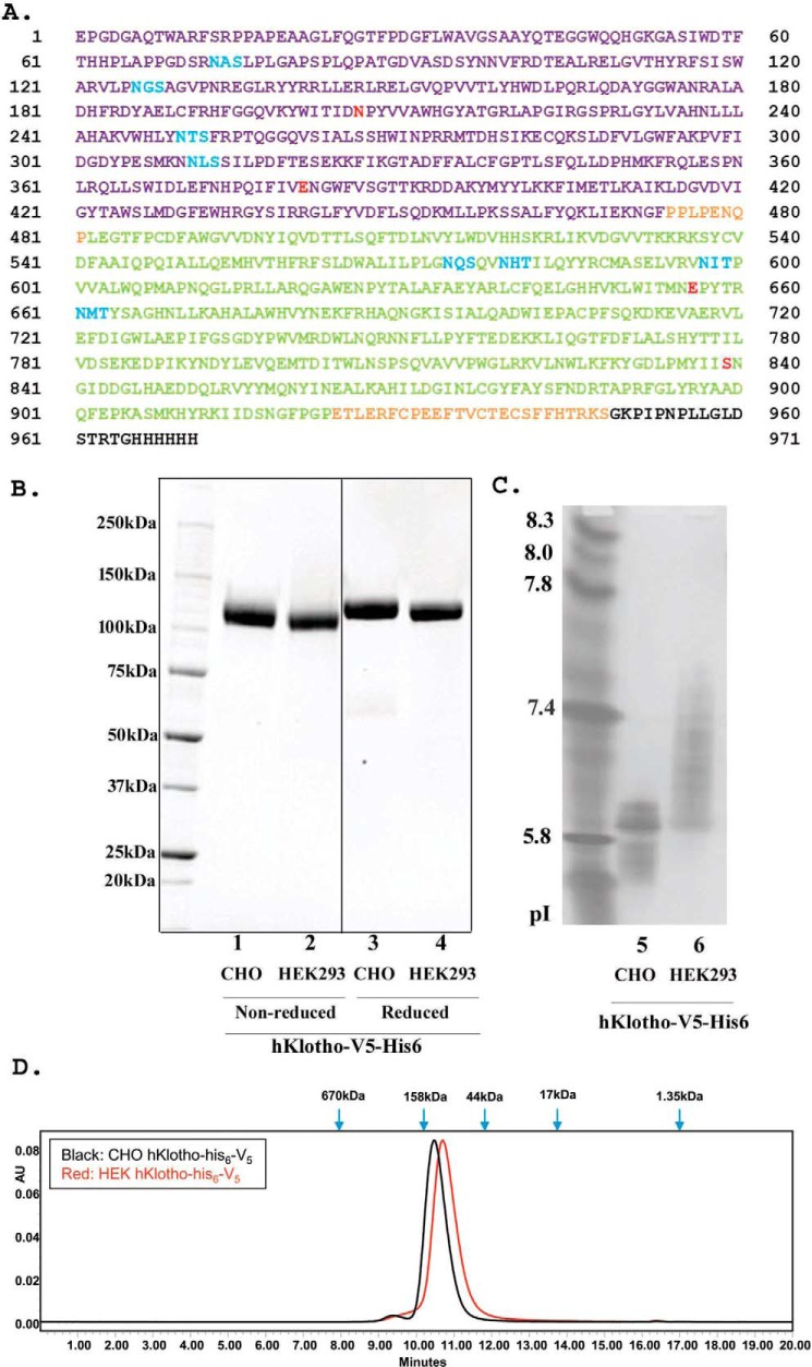
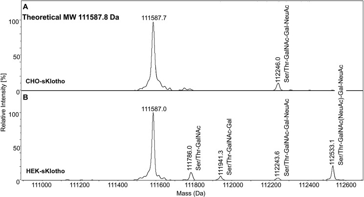
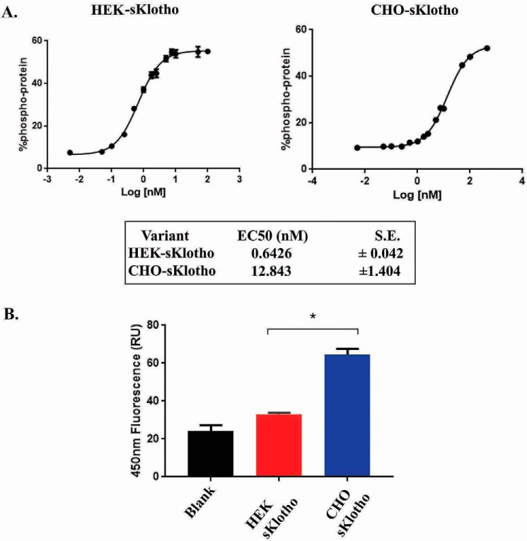
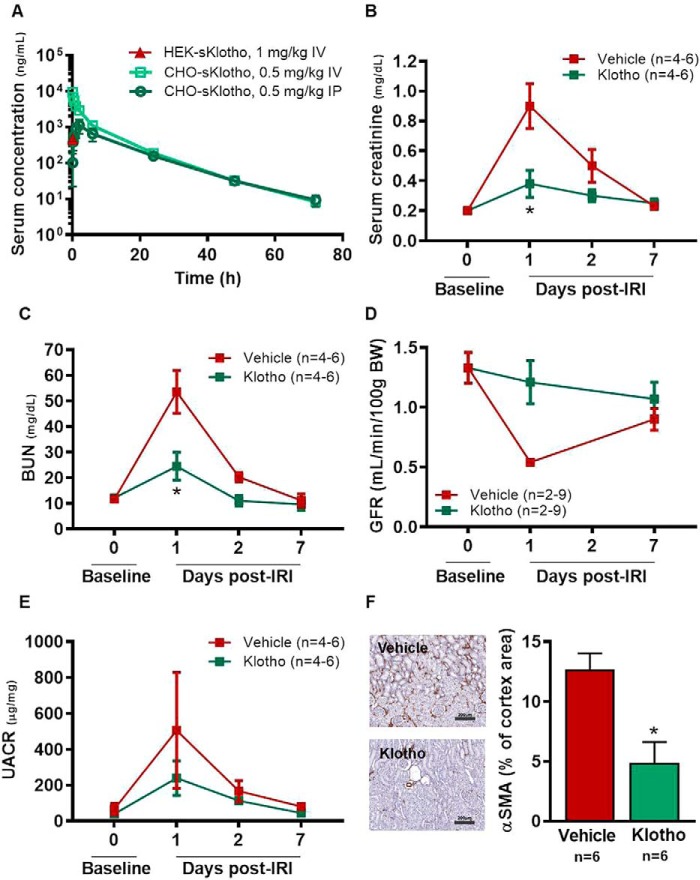
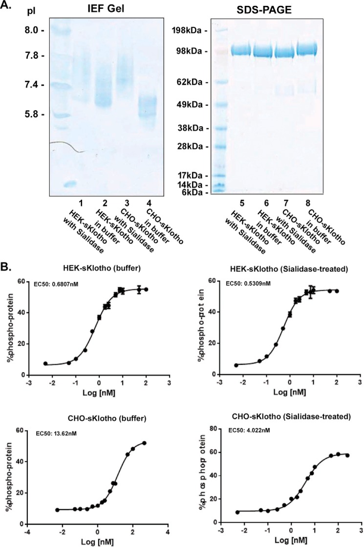
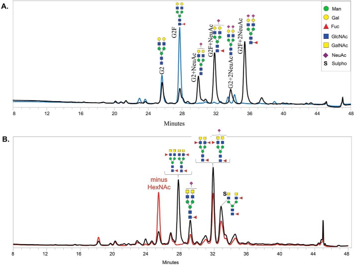
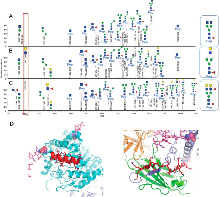
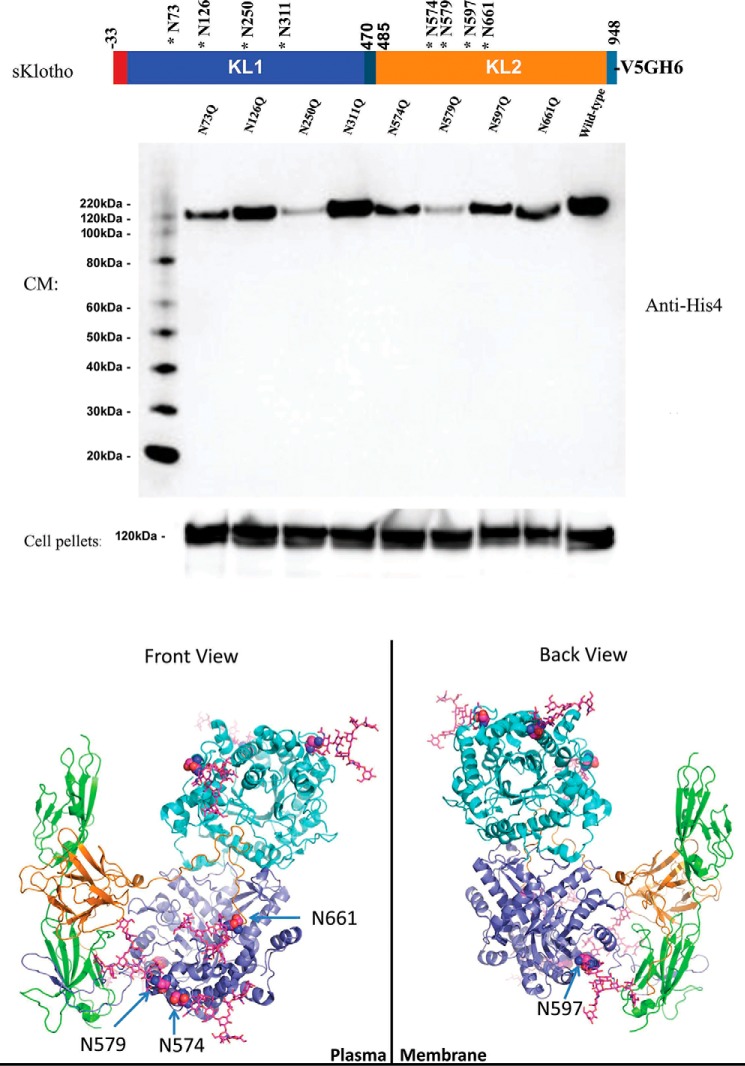
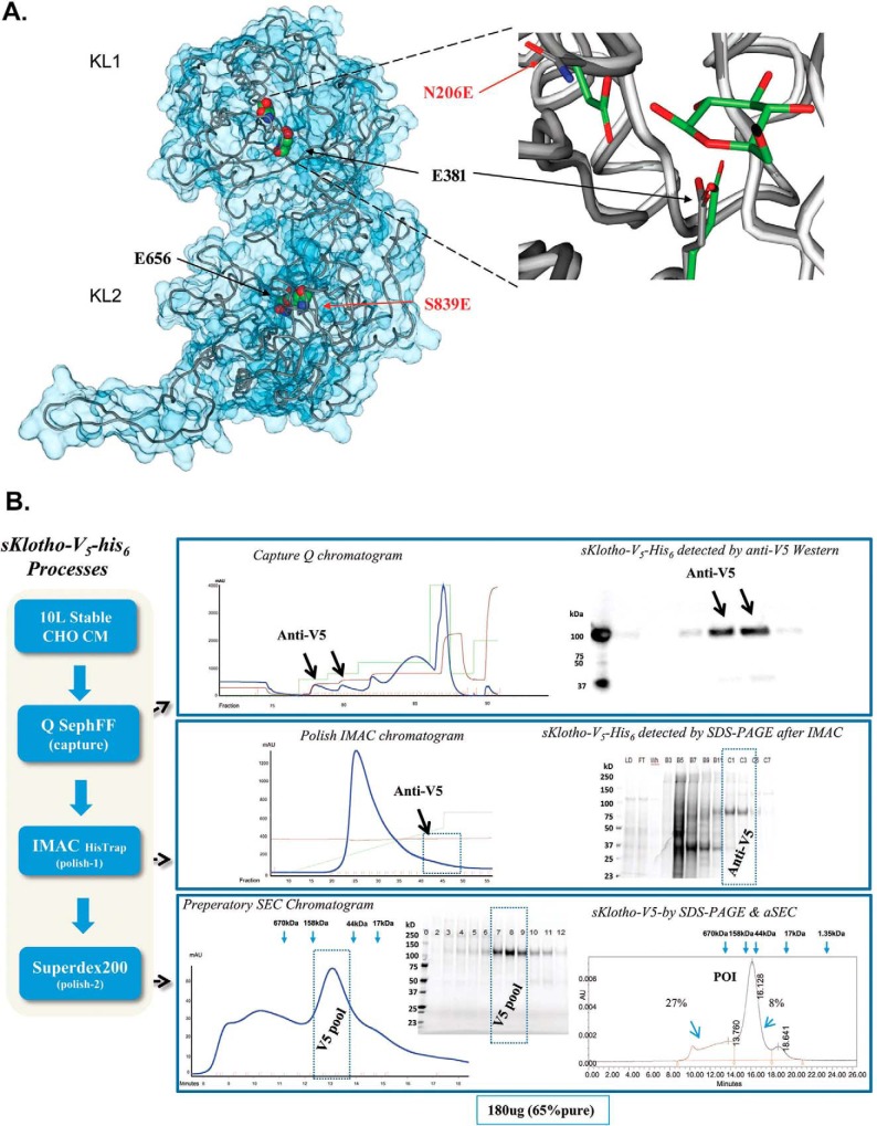
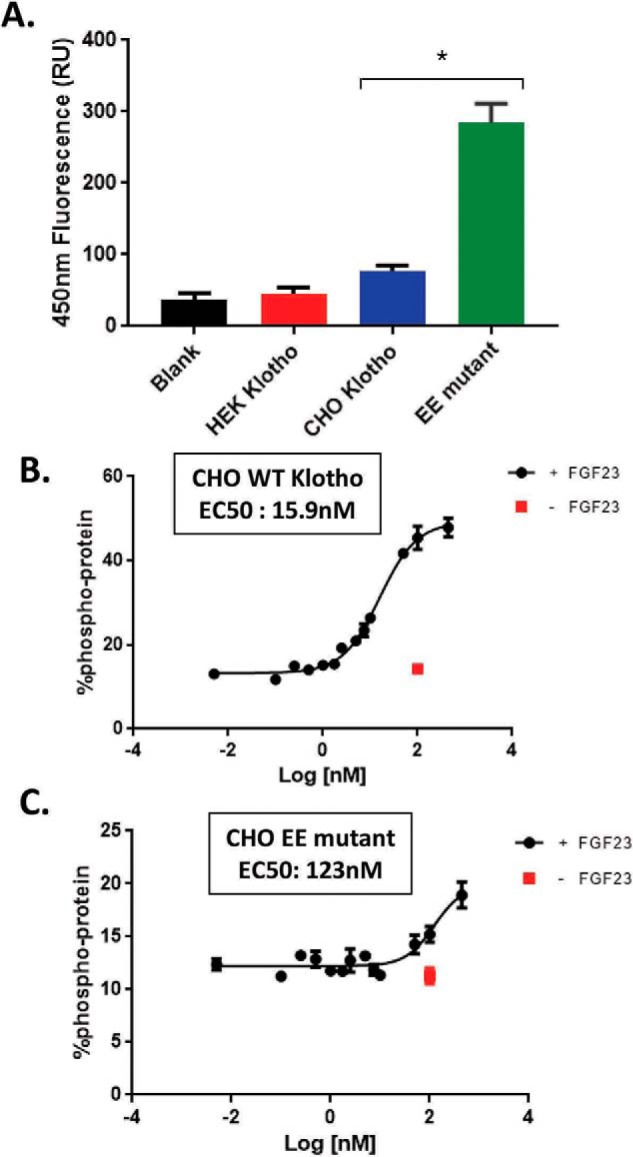
The fortuitously discovered antiaging membrane protein αKlotho (Klotho) is highly expressed in the kidney, and deletion of the Klotho gene in mice causes a phenotype strikingly similar to that of chronic kidney disease (CKD). Klotho functions as a co-receptor for fibroblast growth factor 23 (FGF23) signaling, whereas its shed extracellular domain, soluble Klotho (sKlotho), carrying glycosidase activity, is a humoral factor that regulates renal health. Low sKlotho in CKD is associated with disease progression, and sKlotho supplementation has emerged as a potential therapeutic strategy for managing CKD. Here, we explored the structure-function relationship and post-translational modifications of sKlotho variants to guide the future design of sKlotho-based therapeutics. Chinese hamster ovary (CHO)- and human embryonic kidney (HEK)-derived WT sKlotho proteins had varied activities in FGF23 co-receptor and β-glucuronidase assays in vitro and distinct properties in vivo Sialidase treatment of heavily sialylated CHO-sKlotho increased its co-receptor activity 3-fold, yet it remained less active than hyposialylated HEK-sKlotho. MS and glycopeptide-mapping analyses revealed that HEK-sKlotho is uniquely modified with an unusual N-glycan structure consisting of N,N'-di-N-acetyllactose diamine at multiple N-linked sites, one of which at Asn-126 was adjacent to a putative GalNAc transfer motif. Site-directed mutagenesis and structural modeling analyses directly implicated N-glycans in Klotho's protein folding and function. Moreover, the introduction of two catalytic glutamate residues conserved across glycosidases into sKlotho enhanced its glucuronidase activity but decreased its FGF23 co-receptor activity, suggesting that these two functions might be structurally divergent. These findings open up opportunities for rational engineering of pharmacologically enhanced sKlotho therapeutics for managing kidney disease.

Keywords: Klotho; LacdiNAc; acute kidney injury; fibroblast growth factor receptor (FGFR); glycosidase; glycosylation; mammalian cell expression; pharmacokinetics; sialic acid; signal transduction.


Conflict of interest statement

The authors declare that they have no conflicts of interest with the contents of this article.

Figures

Figure 1.
Figure 1.
Production of a secreted full-length extracellular domain of Klotho in mammalian cells. A, amino acid sequence of a secreted full-length ectodomain of human Klotho. The KL1 domain is shown in violet, and the KL2 domain is green. Putative N-linked sites are blue. V5 tag and His6 tag are black. Four residues at highly conserved positions are red. B, SDS-PAGE analysis of purified sKlotho protein expressed in mammalian cells. sKlotho protein in A was expressed in either stable CHO or transient HEK293 and purified as described under “Experimental procedures.” A spliced region in the gel is indicated with a heavy line. C, IEF gel analysis of purified sKlotho protein. D, SEC analysis of purified sKlotho protein. Bio-Rad gel filtration standards indicated by blue arrows are thyroglobulin (bovine, 670 kDa), γ-globulin (bovine, 158 kDa), ovalbumin (chicken, 44 kDa), myoglobin (horse, 17 kDa), and vitamin B12 (1.35 kDa).
Figure 2.
Figure 2.
Mass spectrometry analysis of CHO-sKlotho (A) and HEK-sKlotho (B). CHO-sKlotho and HEK-sKlotho were digested with PNGase F and subjected to ultrahigh-resolution MS analysis as described under “Experimental procedures.” Structures of mucin-type core-1 O-linked glycans (monosialylated core-1, disialylated core-1) are depicted in addition to Ser/Thr-GalNAc and Ser/Thr-GalNAc-Gal.
Figure 3.
Figure 3.
In vitro functional characterization of sKlotho produced in mammalian cells. A, sKlotho is active in the FGF23 ERK1/2 activation assay. Purified sKlotho proteins were measured in the ERK1/2 activation assay as described under “Experimental procedures.” The error bars at each data point were calculated from three independent experiments. B, sKlotho possessed weak β-glucuronidase activity. Purified sKlotho proteins were measured in the β-glucuronidase assay as described under “Experimental procedures.” The error bars at each data point were calculated from four independent experiments. Data are shown as mean ± S.E. *, p < 0.05 versus HEK sKlotho, ANOVA.
Figure 4.
Figure 4.
Single-dose administration of CHO-sKlotho is protective against IRI in rats. A, pharmacokinetic profiles of HEK- and CHO-sKlotho; B, IRI, serum creatinine; C, IRI, BUN; D, IRI, GFR; E, IRI, albuminuria; F, IRI, renal cortical α-smooth muscle cell actin (scale bar, 200 μm). Data are shown as mean ± S.E. *, p < 0.05 versus vehicle, ANOVA.
Figure 5.
Figure 5.
Sialidase treatment increased the activity of heavily sialylated CHO-sKlotho by 3-fold but had little effect on the less-sialylated HEK-sKlotho in the FGF23 ERK1/2 activation assay. A, CHO- or HEK-sKlotho proteins (0.5 mg each) were treated or mock-treated with sialidase (500 units; New England Biolabs) overnight at room temperature in G1 buffer, repurified through a nickel-nitrilotriacetic acid column, buffer-exchanged, and concentrated at 0.4 mg/ml in PBS buffer. The protein samples were analyzed by SDS-PAGE or IEF gel. B, sialidase-treated or mock-treated sKlotho proteins were measured in the FGF23 ERK1/2 activation assay as described under “Experimental procedures.” The error bars at each data point were calculated from a triplicated experiment.
Figure 6.
Figure 6.
Released 2-AB–labeled N-glycan analysis of CHO-sKlotho (A) and HEK-sKlotho (B). N-Glycans released from CHO- and HEK-sKlotho were subjected to HILIC-FLD glycan analysis as described under “Experimental procedures” and in Fig. S1. A, CHO-sKlotho is heavily sialylated (black trace) and efficiently desialylated by sialidase A digestion (blue trace). B, HEK-sKlotho was found to contain unique N-glycans with LacdiNAc moieties with both sialyation and sulfation (black trace). Exoglycosidase β-N-acetylhexosaminidasef (New England BioLabs; 16-h incubation at 37 °C prior to chromatography) specifically removed one terminal HexNAc from the LacdiNAc (GalNAcβ1–4GlcNAc)-containing N-glycan structures in various proportions due to interference from terminal fucosylation, sialyation, and sulfation (red trace).
Figure 7.
Figure 7.
A–C, LC-MS/MS spectra from glycopeptide mapping (Asn-126) demonstrated that the diagnostic oxonium ion observed at m/z 407.1668 (z = 1) clearly distinguishes N-glycans without and with the LacdiNAc structural determinant, as shown for CHO-sKlotho (A) and HEK-sKlotho (B and C), respectively. The glycopeptide fragmentation patterns of the CHO-sKlotho precursor ion, m/z 875.7305 (z = 3), in A and the HEK-sKlotho precursor ion, m/z 875.7305 (z = 3), in B resemble two isomeric N-glycans (HexNAc4Man3Fuc1) following positive ion LC-MS/MS analysis via HCD, given the similar fragment ion mass-intensity information; however, the m/z 407 diagnostic oxonium ion provides clear differentiation of LacdiNAc moiety in B. Furthermore, the fragmentation patterns of the HEK-sKlotho precursor ion, m/z 1059.8028 (z = 3), in C confirmed the peptide (Pep) VLPN126GSAGVPNR from Klotho and the covalent N-linked oligosaccharide composition as HexNAc6Man3Fuc2 with the putative N-linked oligosaccharide structure containing LacdiNAc as shown above C (major and minor fragment ions are assigned in Table S3). D, structural modeling of sKlotho. Motif 1 (REGLRYYRRLLERLR) is part of an α-helix (in red, running left to right). Motif 2 (KRLIKVDGVVTKKRK) is part of an extended loop and not in a helix (in red, running left to right).
Figure 8.
Figure 8.
Site mutagenesis and structural modeling analysis further indicated that N-glycans might directly modulate Klotho's protein folding and co-receptor function. A, a single N-linked site mutant of sKlotho was transiently transfected into HEK293 cells as described under “Experimental procedures.” Both conditioned medium (CM) and cell pellets were analyzed by SDS-PAGE and immunoblotted with anti-His4 antibody (Qiagen, Germantown, MD). B, structural modeling of sKlotho/FGFR1c/FGF23 complex. Structure from Protein Data Bank entry 5W21 (32) was modeled with extended sugar chains from Fc glycan in Protein Data Bank entry 5VGP.
Figure 9.
Figure 9.
Production of the enzyme-up EE mutant in which two catalytic glutamic acid residues conserved across the glycosidase family members were introduced into sKlotho. A, modeling of sKlotho EE mutant. The Klotho structure (5W21) (32) with active site residues was rendered in a space-filling model (CPK). Mutated residue numbers are indicated in red. The right part of the image is the overlay of the KLrP (2E9L, white peptide backbone and green side chain/ligand carbons) and KL1 (5W21, gray peptide backbone side-chain carbons) active site. B, purification of sKlotho EE mutant from stable CHO production. The sKlotho EE mutant was expressed in stable CHO cells, and conditioned medium was purified as described under “Experimental procedures”. Bio-Rad gel filtration standards (thyroglobulin (bovine, 670 kDa), γ-globulin (bovine, 158 kDa), ovalbumin (chicken, 44 kDa), myoglobin (horse, 17 kDa), and vitamin B12 (1.35 kDa)) are indicated by blue arrows.
Figure 10.
Figure 10.
The enzyme-up EE mutant sKlotho had a 25-fold higher β-glucuronidase activity than the WT sKlotho but was 8-fold less active than the WT protein in the FGF23 signaling assay. A, EE mutant sKlotho, along with the stable CHO and transient HEK WT protein, was measured in the β-glucuronidase assay as described under “Experimental procedures.” The error bars at each data point were calculated from a triplicated experiment. Data are shown as mean ± S.E. *, p < 0.05 versus CHO Klotho, ANOVA. Stable CHO-WT sKlotho (B) and EE mutant sKlotho (C) were measured in the FGF23 ERK1/2 activation assay as described under “Experimental procedures.” The error bars at each data point were calculated from a triplicated experiment.

References

    1. Coresh J., Selvin E., Stevens L. A., Manzi J., Kusek J. W., Eggers P., Van Lente F., and Levey A. S. (2007) Prevalence of chronic kidney disease in the United States. JAMA 298, 2038–2047 10.1001/jama.298.17.2038 - DOI - PubMed
    1. Levey A. S., Atkins R., Coresh J., Cohen E. P., Collins A. J., Eckardt K. U., Nahas M. E., Jaber B. L., Jadoul M., Levin A., Powe N. R., Rossert J., Wheeler D. C., Lameire N., and Eknoyan G. (2007) Chronic kidney disease as a global public health problem: approaches and initiatives—a position statement from Kidney Disease Improving Global Outcomes. Kidney Int. 72, 247–259 10.1038/sj.ki.5002343 - DOI - PubMed
    1. Radhakrishnan J., Remuzzi G., Saran R., Williams D. E., Rios-Burrows N., Powe N., CDC-CKD Surveillance Team, Brück K., Wanner C., Stel V. S., European CKD Burden Consortium, Venuthurupalli S. K., Hoy W. E., Healy H. G., Salisbury A., et al. (2014) Taming the chronic kidney disease epidemic: a global view of surveillance efforts. Kidney Int. 86, 246–250 10.1038/ki.2014.190 - DOI - PMC - PubMed
    1. Grams M. E., Plantinga L. C., Hedgeman E., Saran R., Myers G. L., Williams D. E., Powe N. R., and CDC CKD Surveillance Team (2011) Validation of CKD and related conditions in existing data sets: a systematic review. Am. J. Kidney Dis. 57, 44–54 10.1053/j.ajkd.2010.05.013 - DOI - PMC - PubMed
    1. Stenvinkel P., and Larsson T. E. (2013) Chronic kidney disease: a clinical model of premature aging. Am. J. Kidney Dis. 62, 339–351 10.1053/j.ajkd.2012.11.051 - DOI - PubMed

MeSH terms