. 2020 Mar 27;295(13):4327-4340.
doi: 10.1074/jbc.RA119.012251. Epub 2020 Jan 31.

Exploring Cryptococcus neoformans capsule structure and assembly with a hydroxylamine-armed fluorescent probe

Affiliations

Exploring Cryptococcus neoformans capsule structure and assembly with a hydroxylamine-armed fluorescent probe

Conor J Crawford et al. J Biol Chem. .

Abstract

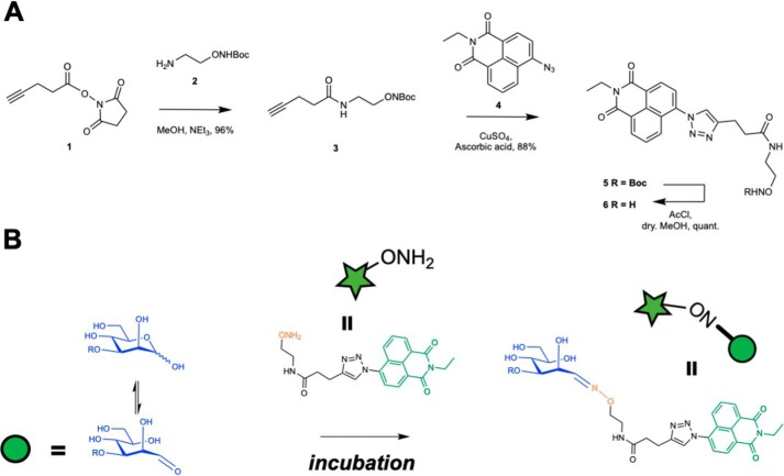
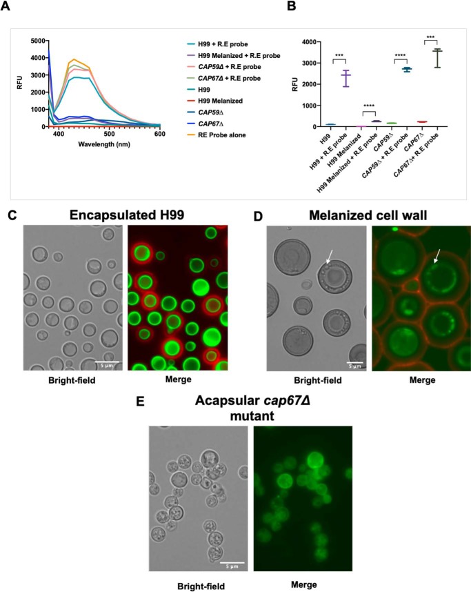
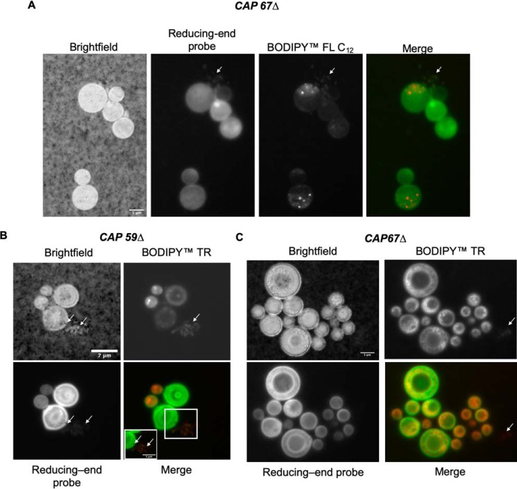
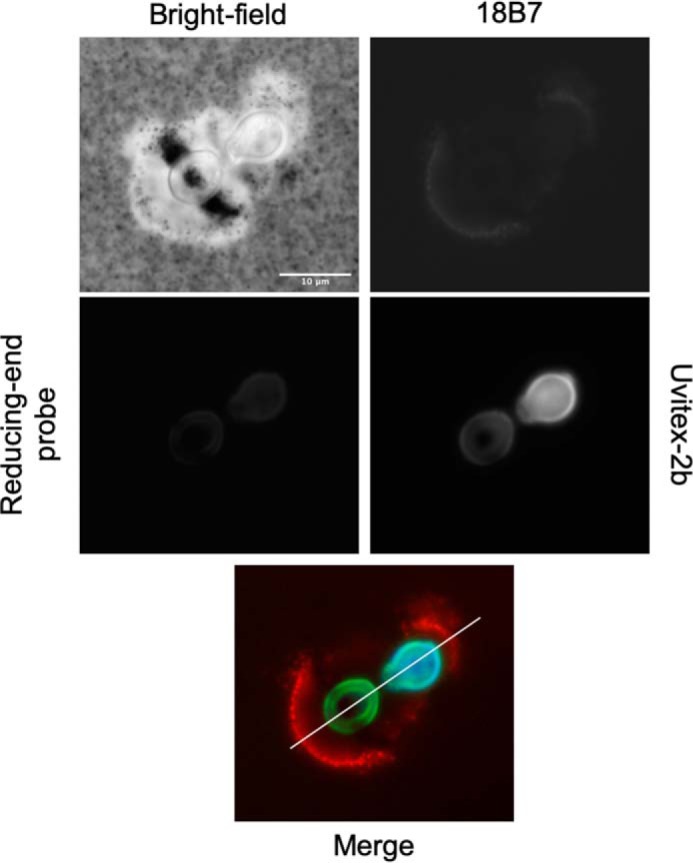
Chemical biology is an emerging field that enables the study and manipulation of biological systems with probes whose reactivities provide structural insights. The opportunistic fungal pathogen Cryptococcus neoformans possesses a polysaccharide capsule that is a major virulence factor, but is challenging to study. We report here the synthesis of a hydroxylamine-armed fluorescent probe that reacts with reducing glycans and its application to study the architecture of the C. neoformans capsule under a variety of conditions. The probe signal localized intracellularly and at the cell wall-membrane interface, implying the presence of reducing-end glycans at this location where the capsule is attached to the cell body. In contrast, no fluorescence signal was detected in the capsule body. We observed vesicle-like structures containing the reducing-end probe, both intra- and extracellularly, consistent with the importance of vesicles in capsular assembly. Disrupting the capsule with DMSO, ultrasound, or mechanical shear stress resulted in capsule alterations that affected the binding of the probe, as reducing ends were exposed and cell membrane integrity was compromised. Unlike the polysaccharides in the assembled capsule, isolated exopolysaccharides contained reducing ends. The reactivity of the hydroxylamine-armed fluorescent probe suggests a model for capsule assembly whereby reducing ends localize to the cell wall surface, supporting previous findings suggesting that this is an initiation point for capsular assembly. We propose that chemical biology is a promising approach for studying the C. neoformans capsule and its associated polysaccharides to unravel their roles in fungal virulence.

Keywords: aminooxy fluorescent probes; biosynthesis; capsule; chemical biology; extracellular vesicles; fungal pathogen; fungi; glucuronoxylomannan; glycobiology; polysaccharide; reducing glycans; vesicles; virulence factor.


Conflict of interest statement

The authors declare that they have no conflicts of interest with the contents of this article

Figures

Figure 1.
Figure 1.
Hydroxylamine-armed fluorescent probe to study the distribution of reducing sugars in C. neoformans. A, synthesis of the R.E probe. B, proposed mechanism of action of the R.E armed fluorescent probe. The R.E probe reacts preferentially with aldehydes to form a stable oxime adduct that can be used to image the localization of reducing ends in the capsule and inside the cell. The hydroxylamine functionality (ONH2) is converted into an oxime (ON) after reacting with the reducing end glycan.
Figure 2.
Figure 2.
Incubation of hydroxylamine-armed fluorescent probe with C. neoformans. A, fluorescence spectrum of C. neoformans cells incubated with and without hydroxylamine-armed probe (R.E probe). Those that have been incubated with the R.E probe have increased emission maxima at ∼453 nm (excitation 360 nm) compared with encapsulated H99, CAP59Δ, or CAP67Δ with the exception of C. neoformans with encapsulated H99 with a melanized cell wall. Melanization of the cell wall is likely quenching the florescence of the probe. B, RFU of C. neoformans cells incubated with the R.E probe are significantly higher at emission maxima (440 nm) of the probe (excitation 360 nm). Error bars represent 95% confidence interval. Statistical significance was determined using unpaired t test (***, p ≤ 0.001; ****, p ≤ 0.0001). Experiments were performed in triplicate. C, encapsulated H99: the capsule shown by anti-GXM Mab 18B7-AF594 (red) displays no reactivity to the R.E probe 6 (green) and is localized at the cell wall–membrane interface. D, melanized C. neoformans cells display the same localization of the R.E probe. The R.E probe appears intracellularly possible in vesicular bodies and displays no reactivity toward the capsule, despite the melanization of the cell wall. E, acapsular mutant C. neoformans cap67Δ incubated with the R.E probe shows bright fluorescence intensity coming from the cytoplasm and the cell wall–membrane interface. Labeling of cytoplasm occurs in spherical vesicle-like structures in acapsular cap67Δ and melanized H99 cells. Scale, 5 μm.
Figure 3.
Figure 3.
Localization of reducing end polysaccharides is maintained across the Cryptococcus genus as labeled by the R.E probe. Incubations with the R.E probe (green), followed by India ink staining allows visualization of the capsule perimeter and where the reducing-ends reside. Any of the Cryptococcal sp. tested resulted in a similar staining pattern, suggesting that capsule architecture and biosynthesis are maintained across species. A, C. neoformans (ATCC 24067). B, C. albidus. C, C. gattii (ATCC 32608). D, C. gattii (ATCC 24065). E, incubation with the R.E probe increases RFU at emission of probe (excitation 360 nm). Error bars represent 95% confidence interval, Statistical significance was determined using unpaired t test (***, p ≤ 0.001; ****, p ≤ 0.0001). Experiments were performed in triplicate. Scale, 5 μm.
Figure 4.
Figure 4.
Capsule perturbation alters probe reactivity. H99 C. neoformans cells were grown in capsule-inducing minimal media for 3 days. Subsequently, the cells were independently processed with three different methods: DMSO, French press, and sonication. Thereafter, the cells were incubated with the fluorescent R.E probe (labels reducing carbohydrates, green) overnight, washed three times, stained for 30 min with Uvitex-2b (stains cell wall chitin, blue), and an 18B7-Alexa Fluor 594 conjugate (stains capsule, red) for 1 h. Scale, 10 μm.
Figure 5.
Figure 5.
Secretion of vesicular bodies in CAP59Δ and CAP67Δ mutants. A, incubation of CAP67Δ with BODIPY FL C12 (lipophilic dye, red) and the R.E probe shows vesicle secretion (arrows). B, CAP59Δ cells incubated with BODIPY TR (lipophilic dye, red) also show signs of secretion of glycan-containing vesicles (inset and arrows). C, CAP67Δ cells incubated with the BODIPY TR and R.E probe show signs of colocalization; however, BODIPY TR also stains nonglycan-containing vesicles (arrows) and shows signs of secretion of nonglycan-containing vesicular bodies (arrow). Scale denoted in bright field: A, 5 μm; B, 7 μm (inset, 5 μm); and C, 5 μm.
Figure 6.
Figure 6.
Dynamic reorganization of C. neoformans polysaccharide capsule and cell–cell wall to facilitate budding. India ink staining perpendicular to emerging bud correlates with reorganization of chitin cell wall and capsule. 18B7 and R.E probe both have been polarized along the budding axis (line merge). This causes a change in capsule density perpendicular to the budding axis allowing India ink staining to penetrate deeper when gentle pressure is applied to the coverslip. Scale, 10 μm.
Figure 7.
Figure 7.
Applying shear pressure to understand probe localization. Application of shear pressure to glass slides caused a population of cells to rupture. Several fragments of cells can be seen (arrows), where the capsule can be seen attached to the cell wall, and also where the R.E probe localization occurs. Scale, 10 μm.
Figure 8.
Figure 8.
Exopolysaccharides of C. neoformans contain reducing ends. A, R.E probe was incubated with CPS, EPS (>10 kDa and >100 kDa), chitin (positive control), or alone for 24 h. The incubation was repeated in three independent experiments. Following dialysis, fluorescence emission spectra were recorded (excitation 360 nm) and compared with their nonincubated controls, showing an increase in fluorescence at the probe's emission maxima, indicating the R.E probe is forming a conjugate with the reducing end of various C. neoformans EPS and CPS isolates. Polysaccharides that were incubated with the R.E probe show >2-fold increase in RFU compared with controls, revealing that the shed exopolysaccharide contains reducing ends. Error bars represent 95% confidence intervals. Statistical significance was determined using unpaired t test (****, p ≤ 0.0001). B, hypothesis: if reducing ends are present in the exopolysaccharide of C. neoformans, they would form stable conjugates with the R.E probe, which could be determined using a fluorescence spectrometer. Glycan notification followed the Symbol Nomenclature for Glycans (SNFG). C, 1H NMR analysis of >10- and >100-kDa exopolysaccharide isolates revealed the presence of aromatic peaks in the region of δ 7.7–8.6 ppm, which are not present in the original EPS fraction. Exploring C. neoformans with hydroxylamine-armed probe 28 aromatic signals are characteristic of the R.E probe. Characteristic structural reporter groups of anomeric mannose protons can be visualized from δ 5.5 to 4.9 ppm.
Figure 9.
Figure 9.
Diagram illustrating three hypotheses for capsule assembly in C. neoformans. Polysaccharide molecules are shown in a linear manner for simplicity, with the localization of the reducing ends a key point for this depiction. The most parsimonious interpretation of the R.E. probe data supports hypothesis 1.

Similar articles

Cited by

References

    1. Park B. J., Wannemuehler K. A., Marston B. J., Govender N., Pappas P. G., and Chiller T. M. (2009) Estimation of the current global burden of cryptococcal meningitis among persons living with HIV/AIDS. AIDS 23, 525–530 10.1097/QAD.0b013e328322ffac - DOI - PubMed
    1. Garcia-Hermoso D., Janbon G., and Dromer F. (1999) Epidemiological evidence for dormant Cryptococcus neoformans infection. J. Clin. Microbiol. 37, 3204–3209 10.1128/JCM.37.10.3204-3209.1999 - DOI - PMC - PubMed
    1. Goldman D. L., Khine H., Abadi J., Lindenberg D. J., Pirofski La., Niang R., and Casadevall A. (2001) Serologic evidence for Cryptococcus neoformans infection in early childhood. Pediatrics 107, E66 10.1542/peds.107.5.e66 - DOI - PubMed
    1. Singh N., Dromer F., Perfect J. R., and Lortholary O. (2008) Cryptococcosis in solid organ transplant recipients: current state of the science. Clin. Infect. Dis. 47, 1321–1327 10.1086/592690 - DOI - PMC - PubMed
    1. Martinez L. R., and Casadevall A. (2015) Biofilm formation by Cryptococcus neoformans. Microbiol. Spectr. 3, 10.1128/microbiolspec.MB-0006-2014 - DOI - PubMed

Publication types

MeSH terms

LinkOut - more resources