. 2020 Mar 13;295(11):3734-3745.
doi: 10.1074/jbc.RA119.011322. Epub 2020 Jan 31.

Functional analysis of Clostridium difficile sortase B reveals key residues for catalytic activity and substrate specificity

Affiliations

Functional analysis of Clostridium difficile sortase B reveals key residues for catalytic activity and substrate specificity

Chia-Yu Kang et al. J Biol Chem. .

Abstract

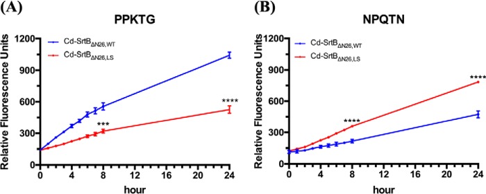
Most of Gram-positive bacteria anchor surface proteins to the peptidoglycan cell wall by sortase, a cysteine transpeptidase that targets proteins displaying a cell wall sorting signal. Unlike other bacteria, Clostridium difficile, the major human pathogen responsible for antibiotic-associated diarrhea, has only a single functional sortase (SrtB). Sortase's vital importance in bacterial virulence has been long recognized, and C. difficile sortase B (Cd-SrtB) has become an attractive therapeutic target for managing C. difficile infection. A better understanding of the molecular activity of Cd-SrtB may help spur the development of effective agents against C. difficile infection. In this study, using site-directed mutagenesis, biochemical and biophysical tools, LC-MS/MS, and crystallographic analyses, we identified key residues essential for Cd-SrtB catalysis and substrate recognition. To the best of our knowledge, we report the first evidence that a conserved serine residue near the active site participates in the catalytic activity of Cd-SrtB and also SrtB from Staphylococcus aureus The serine residue indispensable for SrtB activity may be involved in stabilizing a thioacyl-enzyme intermediate because it is neither a nucleophilic residue nor a substrate-interacting residue, based on the LC-MS/MS data and available structural models of SrtB-substrate complexes. Furthermore, we also demonstrated that residues 163-168 located on the β6/β7 loop of Cd-SrtB dominate specific recognition of the peptide substrate PPKTG. The results of this work reveal key residues with roles in catalysis and substrate specificity of Cd-SrtB.

Keywords: Clostridium difficile; crystal structure; cysteine transpeptidase; enzyme catalysis; fluorescence resonance energy transfer (FRET); protein chemistry; protein purification; protein sorting; protein structure; sortase B; substrate specificity.


Conflict of interest statement

The authors declare that they have no conflicts of interest with the contents of this article

Figures

Figure 1.
Figure 1.
Effect of mutation at residue Ser-207 on the enzymatic activity of Cd-SrtBΔN26 assessed by FRET-based assay. A, purified Cd-SrtBΔN26,WT and Cd-SrtBΔN26,S207A (120 μm) were incubated with PPKTG-containing peptide (20 μm) at 37 °C. The fluorescence signals were measured every hour during the first 8 h and at 24 h. B, the ratios of fluorescence units of Cd-SrtBΔN26,S207A/Cd-SrtBΔN26 at 24 h are shown. C, purified Cd-SrtBΔN26,WT, Cd-SrtBΔN26,S207A, Cd-SrtBΔN26,C209A, and Cd-SrtBΔN26,S207A C209A (120 μm) were incubated with PPKTG-containing peptide (20 μm) at 37 °C. The fluorescence signals were measured every hour during the first 8 h and at 24 h. D, the ratios of fluorescence units Cd-SrtBΔN26,S207A/Cd-SrtBΔN26,WT, Cd-SrtBΔN26,C209A/Cd-SrtBΔN26,WT, and Cd-SrtBΔN26,S207A C209A/Cd-SrtBΔN26,WT at 24 h are shown. The data represent the means ± S.D. of three independent experiments. **, p < 0.01; ****, p < 0.0001; unpaired t test.
Figure 2.
Figure 2.
LC-MS/MS analysis for peptide cleavage site. A, MS extracted ion chromatographs (EIC) are shown for cleavage of Dabcyl-PVPPKTGDSTTIIGE-Edans by Cd-SrtBΔN26,WT (upper panel) and Cd-SrtBΔN26,C209A (lower panel). B, MS/MS spectra of the cleaved substrate peptide from the reaction mixture of Cd-SrtBΔN26,WT with PPKTG-containing peptide shows a doubly charged ion at m/z 570.7421 for MH22+ corresponding to the mass of the cleaved peptide product GDSTTIIGE-Edans. C, the MS/MS spectrum shows a doubly charged ion at m/z 730.8409 for MH22+ corresponding to the mass of the scrambled peptide PVGSSTPDSTTIIGE incubated with Cd-SrtBΔN26,WT. The labeled peaks correspond to masses of y and b ions of the peptide.
Figure 3.
Figure 3.
Crystal structure of the Cd-SrtBΔN26,S207A and structural comparison with Cd-SrtBΔN26,WT. A, overall crystal structure of Cd-SrtBΔN26,S207A is shown in blue (left panel). The β6/β7 loop region (residues 162–167) and the β6/β7 loop region (residues 210–216) are disordered in this structure. A zooming structure of Cd-SrtBΔN26,S207A around the residue Ser-207 with a FoFc omit electron density map at the 2 σ level is shown in gray chicken wire (right panel). B, superposition of Cd-SrtBΔN26,WT (PDB 5GYJ, yellow) and Cd-SrtBΔN26,S207A (PDB 6KYC, blue) yields an RMSD value of 0.174 for 176 Cα atoms. The side chains of Ser-207 in Cd-SrtBΔN26,WT and Ala-207 of Cd-SrtBΔN26,S207A are shown as sticks on β7-strand.
Figure 4.
Figure 4.
Mutational effects at residues Cys-194 and Ser-192 on enzymatic activity of Sa-SrtBΔN29. A, the fluorescence signals were monitored after incubating the purified Sa-SrtBΔN29,WT, Sa-SrtBΔN26,C194A, Sa-SrtBΔN29,S192A, and Sa-SrtBΔN29,S192A C194A (120 μm) with NPQTN-containing peptide (20 μm), respectively, at 37 °C for every hour during the first 8 h and at 24 h. B, the relative fluorescence signals of Sa-SrtBΔN26,C194A, Sa-SrtBΔN29,S192A, and Sa-SrtBΔN29, S192A C194A are significantly lower than WT Sa-SrtBΔN29 at 24 h. The data represent the means ± S.D. of three independent experiments. ****, p < 0.0001; unpaired t test.
Figure 5.
Figure 5.
Effects of mutation at His-116 and Arg-217 on enzymatic activity of Cd-SrtB. A, purified Cd-SrtBΔN26,WT, Cd-SrtBΔN26,C209A, Cd-SrtBΔN26,R217A, and Cd-SrtBΔN26,H116A (120 μm) were incubated with PPKTG-containing peptide (20 μm), respectively, at 37 °C. The fluorescence signals were measured every hour during the first 8 h and at 24 h. B, the ratios of fluorescence units of Cd-SrtBΔN26,C209A/Cd-SrtBΔN26,WT, Cd-SrtBΔN26,R217A/Cd-SrtBΔN26,WT, and Cd-SrtBΔN26,H116A/Cd-SrtBΔN26,WT at 24 h are shown. The data represent the means ± S.D. of three independent experiments. n.s., nonsignificant. **, p < 0.01; ****, p < 0.0001; unpaired t test.
Figure 6.
Figure 6.
Crystal structure of Cd-SrtBΔN26,R17A and structural comparison with WT Cd-SrtBΔN26,WT. A, overall crystal structure of Cd-SrtBΔN26,R217A is displayed in green (left panel). The β6/β7 loop region (residues 162–167) and the β6/β7 loop region (residues 210–216) are disordered in this structure. A zooming structure of Cd-SrtBΔN26,R217A around residue Arg-217 with a FoFc omit electron density map at the 2 σ level is shown in gray chicken wire (right panel). B, superposition of Cα atoms on the structure of WT Cd-SrtBΔN26,WT (PDB code 5GYJ) and Cd-SrtBΔN26,R217A are presented in yellow and green, with RMSD = 0.239 for 176 Cα atoms, respectively. The side chains of Arg-217 of Cd-SrtBΔN26,WT and Ala217 of Cd-SrtBΔN26,R217A are shown as sticks on the β8-strand.
Figure 7.
Figure 7.
Substrate specificity of Cd-SrtBΔN26,WT and Sa-SrtBΔN29,WT. The fluorescence signals were monitored to calculate the enzymatic activity of Cd-SrtBΔN26,WT and Sa-SrtBΔN29,WT for every hour during the first 8 h and at 24 h at 37 °C with 20 μm peptide substrate PPKTG (A) or NPQTN (B). The data represent the means ± S.D. of three independent experiments. **, p < 0.01; ***, p < 0.001; unpaired t-test.
Figure 8.
Figure 8.
Effect of β6/β7 loop substitution on the enzymatic activity of Cd-SrtBΔN26,WT by FRET-based assay. Residues 163–168 on β6/β7 loop in Cd-SrtBΔN26,WT was replaced with the corresponding residues 177–182 from Sa-SrtB β6/β7 loop. Purified Cd-SrtBΔN26,LS was incubated with a peptide containing PPKTG motif (A) or NPQTN motif (B). The fluorescence signals were measured every hour during the first 8 h and at 24 h at 37 °C. The data represent the means ± S.D. of three independent experiments. ***, p < 0.001; ****, p < 0.0001; unpaired t test.

Similar articles

Cited by

References

    1. Peery A. F., Dellon E. S., Lund J., Crockett S. D., McGowan C. E., Bulsiewicz W. J., Gangarosa L. M., Thiny M. T., Stizenberg K., Morgan D. R., Ringel Y., Kim H. P., DiBonaventura M. D., Carroll C. F., Allen J. K., et al. (2012) Burden of gastrointestinal disease in the United States: 2012 update. Gastroenterology 143, 1179–1187.e3 10.1053/j.gastro.2012.08.002 - DOI - PMC - PubMed
    1. Asempa T. E., and Nicolau D. P. (2017) Clostridium difficile infection in the elderly: an update on management. Clin. Interv. Aging 12, 1799–1809 10.2147/CIA.S149089 - DOI - PMC - PubMed
    1. Garey K. W., Sethi S., Yadav Y., and DuPont H. L. (2008) Meta-analysis to assess risk factors for recurrent Clostridium difficile infection. J. Hosp. Infect. 70, 298–304 10.1016/j.jhin.2008.08.012 - DOI - PubMed
    1. Honda H., Yamazaki A., Sato Y., and Dubberke E. R. (2014) Incidence and mortality associated with Clostridium difficile infection at a Japanese tertiary care center. Anaerobe 25, 5–10 10.1016/j.anaerobe.2013.10.004 - DOI - PubMed
    1. Johnson S. (2009) Recurrent Clostridium difficile infection: a review of risk factors, treatments, and outcomes. J. Infect. 58, 403–410 10.1016/j.jinf.2009.03.010 - DOI - PubMed

Publication types

MeSH terms

LinkOut - more resources