. 2020 Mar 16;7(2):ENEURO.0453-19.2020.
doi: 10.1523/ENEURO.0453-19.2020. Print 2020 Mar/Apr.

Muscarinic Modulation of SK2-Type K+ Channels Promotes Intrinsic Plasticity in L2/3 Pyramidal Neurons of the Mouse Primary Somatosensory Cortex

Affiliations

Muscarinic Modulation of SK2-Type K+ Channels Promotes Intrinsic Plasticity in L2/3 Pyramidal Neurons of the Mouse Primary Somatosensory Cortex

Daniel F Gill et al. eNeuro. .

Abstract

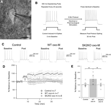
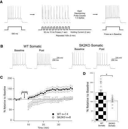
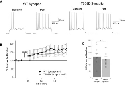
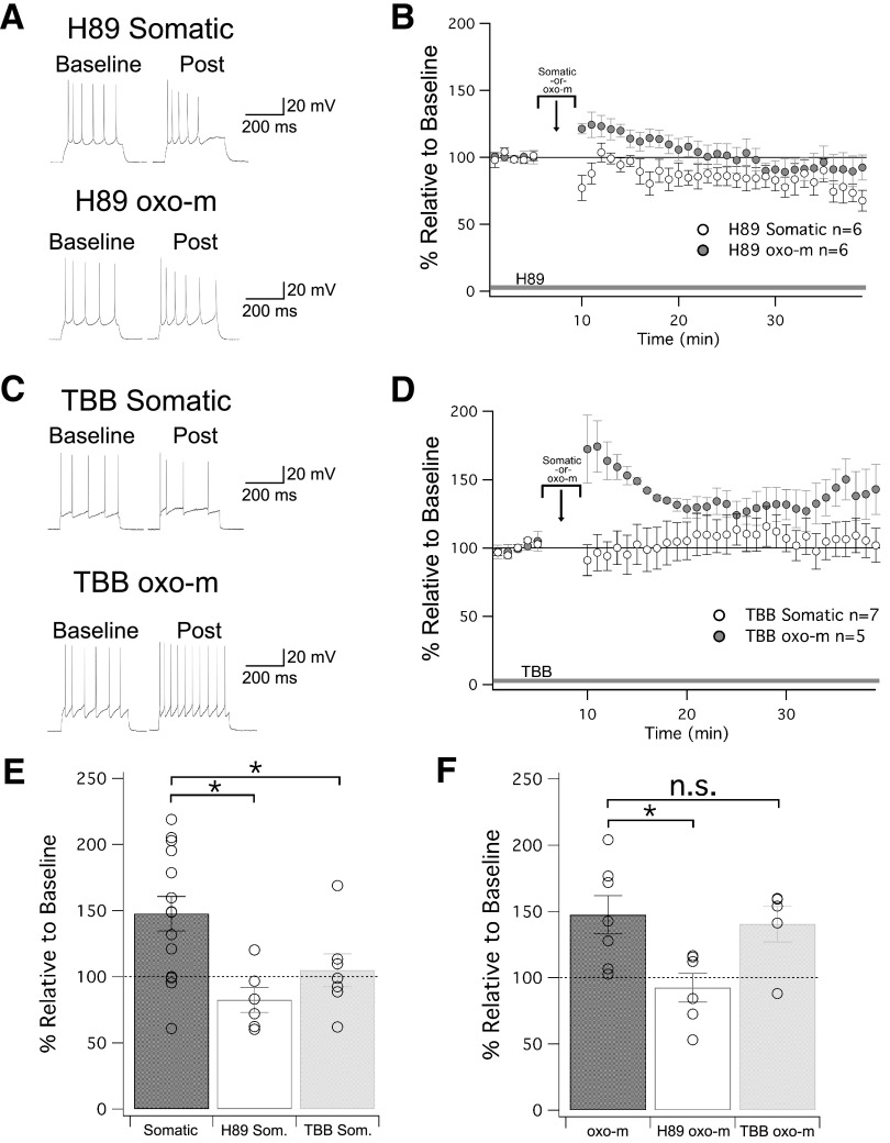
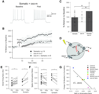
Muscarinic acetylcholine receptors (mAChRs) inhibit small-conductance calcium-activated K+ channels (SK channels) and enhance synaptic weight via this mechanism. SK channels are also involved in activity-dependent plasticity of membrane excitability ("intrinsic plasticity"). Here, we investigate whether mAChR activation can drive SK channel-dependent intrinsic plasticity in L2/3 cortical pyramidal neurons. Using whole-cell patch-clamp recordings from these neurons in slices prepared from mouse primary somatosensory cortex (S1), we find that brief bath application of the mAChR agonist oxotremorine-m (oxo-m) causes long-term enhancement of excitability in wild-type mice that is not observed in mice deficient of SK channels of the SK2 isoform. Similarly, repeated injection of depolarizing current pulses into the soma triggers intrinsic plasticity that is absent from SK2 null mice. Intrinsic plasticity lowers spike frequency adaptation and attenuation of spike firing upon prolonged activation, consistent with SK channel modulation. Depolarization-induced plasticity is prevented by bath application of the protein kinase A (PKA) inhibitor H89, and the casein kinase 2 (CK2) inhibitor TBB, respectively. These findings point toward a recruitment of two known signaling pathways in SK2 regulation: SK channel trafficking (PKA) and reduction of the calcium sensitivity (CK2). Using mice with an inactivation of CaMKII (T305D mice), we show that intrinsic plasticity does not require CaMKII. Finally, we demonstrate that repeated injection of depolarizing pulses in the presence of oxo-m causes intrinsic plasticity that surpasses the plasticity amplitude reached by either manipulation alone. Our findings show that muscarinic activation enhances membrane excitability in L2/3 pyramidal neurons via a downregulation of SK2 channels.

Keywords: engram; ensemble; excitability; learning; neocortex; pyramidal cell.


Figures

Figure 1.
Figure 1.
Muscarinic signaling induces changes in intrinsic excitability through SK2 channels. A, Differential interference contrast (DIC) image of patch-clamp recording from a layer 2/3 pyramidal neuron in a slice prepared from S1 cortex; scale bar = 10 μm. B, Test protocol for intrinsic plasticity. A stable period of 5 min of baseline was collected, where 500-ms pulses were delivered at 0.05 Hz, eliciting four to eight spikes. Following this is a 5-min induction protocol, where oxo-m (or electrical stimulation in later figures) is applied to the slice. Following induction, cells are monitored with the same test pulses used in baseline. C, Example traces of baseline and postinduction for ACSF wash-on (control), oxo-m wash-on, and oxo-m wash-on in SK2KO cells. D, Time graph for changes in spiking relative to baseline for all three groups. oxo-m/ACSF wash on occurs from minute 5 to 10. E, Bar graph of each groups change in spiking relative to baseline. oxo-m significantly increased from baseline (p = 0.017), while ACSF and oxo-m in SK2KO mice did not (p = 0.68 and p = 0.57, respectively). Additionally, the increase observed in oxo-m was significantly greater than the changes observed in the ACSF and SK2KO oxo-m groups (p = 0.021 and p = 0.042, respectively). *p < 0.05.
Figure 2.
Figure 2.
Induction of intrinsic plasticity through somatic depolarization is SK2 dependent. A, Protocol for inducing changes in intrinsic plasticity, modeled off of Mahon and Charpier (2012). As before, a test pulse of 500 ms is repeated at 0.05 Hz in order to induce four to eight spikes. Following a 5-min baseline, the induction protocol injects depolarizing current at 10 Hz (50-ms pulses), evoking one to three spikes per pulse for 1 s, followed by 2 s of holding current. This 3-s sequence is repeated 100 times for a total of 5 min, and then test pulses identical to baseline are again delivered to monitor for changes in excitability. B, Example traces of WT neurons and SK2KO neurons before and after the induction protocol. C, Time graph for changes in spiking relative to baseline, induction protocol occurs from minute 5 to 10. D, Bar graph for change in spiking relative to baseline. WT significantly increased from baseline, while SK2KO did not (p = 0.0028 and p = 0.55, respectively). The increase observed in WT cells was significantly greater than the changes observed in the SK2KO group (p = 0.044). *p < 0.05.
Figure 3.
Figure 3.
Induction of intrinsic plasticity is CaMKII independent. A, Example traces of WT neurons and T305D neurons before and after the induction protocol. The induction protocol was modified from somatic depolarization where an extracellular stimulus was used in lieu of somatic stimulation (further details in text). B, Time graph for changes in spiking relative to baseline, induction protocol occurs from minute 5 to 10. C, Bar graph for change in spiking relative to baseline. WT and T305D both significantly increased from baseline (p = 0.048 and p = 0.040, respectively). The comparative increase between these two groups was not significantly different (p = 0.27). n.s. = nonsignificant.
Figure 4.
Figure 4.
The role of PKA and CK2 in regulating SK2 channels in intrinsic plasticity. A, Example traces for cells treated with H89, a PKA inhibitor that was present in the bath for the duration of the experiment. B, Time graph for changes in spiking relative to baseline. Either somatic depolarization or oxo-m was applied from minute 5 to 10. C, Example traces for cells treated with TBB, a CK2 inhibitor that was present in the bath for the duration of the experiment. D, Time graph for changes in spiking relative to baseline. Either somatic depolarization or oxo-m was applied from minute 5 to 10. E, Bar graph for depolarization-induced changes in spiking relative to baseline. Both H89 and TBB bath application prevented intrinsic plasticity. F, Bar graph for oxo-m-induced changes in spiking relative to baseline. H89, but not TBB, prevented intrinsic plasticity; *p < 0.05. n.s. = nonsignificant.
Figure 5.
Figure 5.
Intersection of somatic and muscarinic activation. A, Example trace of a cell that received somatic depolarization while oxo-m was in the bath. B, Time graph for changes in spiking relative to baseline, somatic, and oxo-m stimulation occurs at minute 5 to 10. C, Bar graph for change in spiking relative to baseline. Combined stimulation was significantly different from baseline (p = 4.9 × 10⁻⁴), and significantly different from somatic or oxo-m stimulation alone (p = 0.035 and p = 0.044, respectively). D, Diagram for how somatic depolarization and muscarinic pathways overlap and interact with SK2 channels. E, Difference in initial firing rate for somatic and synaptic induction protocols. Both groups increased their firing rate per sweep from baseline to post (p = 0.002 and p = 0.020, respectively). F, Spike attenuation ratios for somatic and synaptic induction protocols. The spike attenuation ratio is a ratio of the spiking that takes place in the first half of the sweep, and decreases for both somatic and synaptic cell groups (p = 0.045 and p = 0.034, respectively). G, Shift in attenuation ratio is strongly correlated to change in intrinsic excitability. All cells from groups which had ACSF in the bath during baseline and post are plotted, indicating a strong connection between firing later in sweeps and intrinsic plasticity (p = 3.8 × 10⁻⁴).

Similar articles

Cited by

References

    1. Agmon A, Connors BW (1991) Thalamocortical responses of mouse somatosensory (barrel) cortex in vitro. Neuroscience 41:365–379. 10.1016/0306-4522(91)90333-j - DOI - PubMed
    1. Allen D, Fakler B, Maylie J, Adelman JP (2007) Organization and regulation of small conductance Ca2+ -activated K+ channel multiprotein complexes. J Neurosci 27:2369–2376. 10.1523/JNEUROSCI.3565-06.2007 - DOI - PMC - PubMed
    1. Bear MF, Singer W (1986) Modulation of visual cortical plasticity by acetylcholine and noradrenaline. Nature 320:172–176. 10.1038/320172a0 - DOI - PubMed
    1. Belmeguenai A, Hosy E, Bengtsson F, Pedroarena CM, Piochon C, Teuling E, He Q, Ohtsuki G, De Jeu MT, Elgersma Y, De Zeeuw CI, Jörntell H, Hansel C (2010) Intrinsic plasticity complements long-term potentiation in parallel fiber input gain control in cerebellar Purkinje cells. J Neurosci 30:13630–13643. 10.1523/JNEUROSCI.3226-10.2010 - DOI - PMC - PubMed
    1. Betterton RT, Broad LM, Tsaneva-Atanasova K, Mellor JR (2017) Acetylcholine modulates gamma frequency oscillations in the hippocampus by activation of muscarinic M1 receptors. Eur J Neurosci 45:1570–1585. 10.1111/ejn.13582 - DOI - PMC - PubMed

Publication types

Substances

LinkOut - more resources