. 2020 Jan 31;11(1):633.
doi: 10.1038/s41467-020-14349-2.

Preclinical development of a miR-132 inhibitor for heart failure treatment

Affiliations

Preclinical development of a miR-132 inhibitor for heart failure treatment

Ariana Foinquinos et al. Nat Commun. .

Abstract

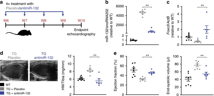
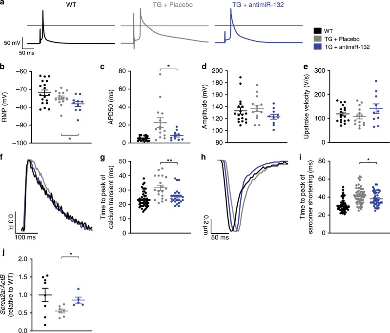
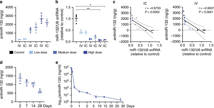
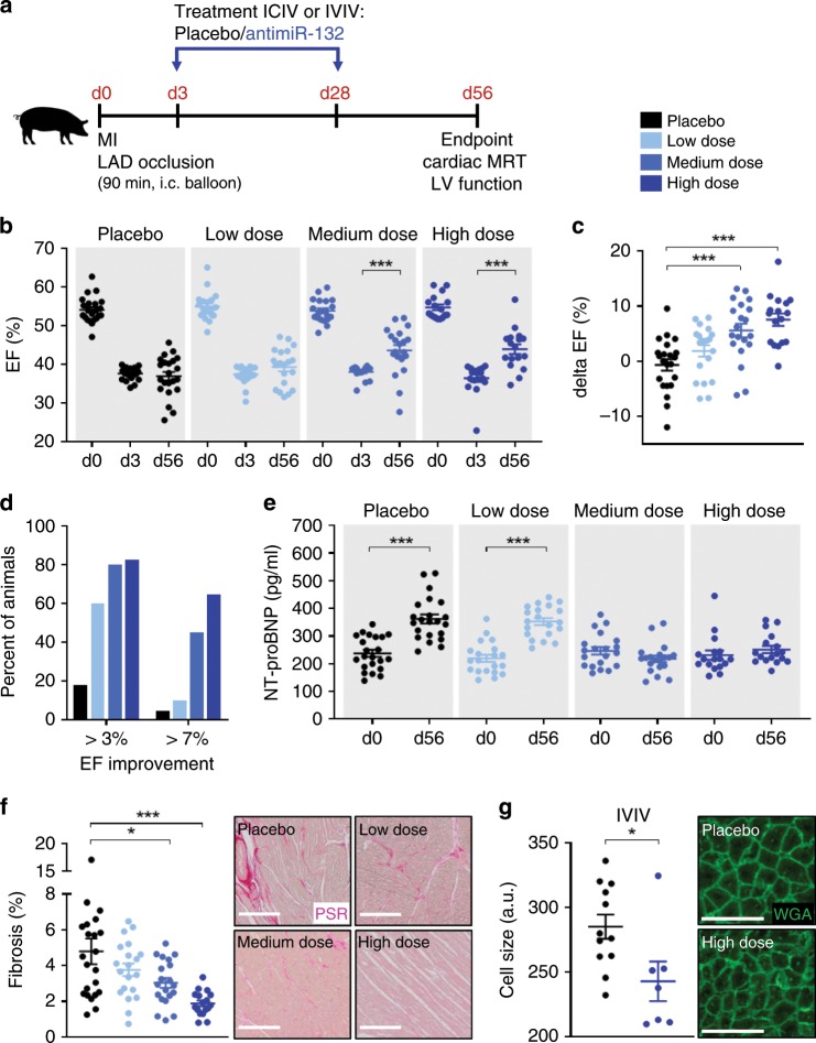
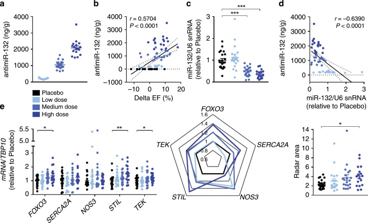
Despite proven efficacy of pharmacotherapies targeting primarily global neurohormonal dysregulation, heart failure (HF) is a growing pandemic with increasing burden. Treatments mechanistically focusing at the cardiomyocyte level are lacking. MicroRNAs (miRNA) are transcriptional regulators and essential drivers of disease progression. We previously demonstrated that miR-132 is both necessary and sufficient to drive the pathological cardiomyocytes growth, a hallmark of adverse cardiac remodelling. Therefore, miR-132 may serve as a target for HF therapy. Here we report further mechanistic insight of the mode of action and translational evidence for an optimized, synthetic locked nucleic acid antisense oligonucleotide inhibitor (antimiR-132). We reveal the compound's therapeutic efficacy in various models, including a clinically highly relevant pig model of HF. We demonstrate favourable pharmacokinetics, safety, tolerability, dose-dependent PK/PD relationships and high clinical potential for the antimiR-132 treatment scheme.


Conflict of interest statement

S.B. and T.T. are co-founders and hold shares of Cardior Pharmaceuticals GmbH. T.T., S.B. and A.F. filed and licensed patents through the Hannover Medical School to Cardior Pharmaceuticals GmbH. T.T., S.B., S.R., C.G. and J.V. are currently part or fulltime employees of Cardior Pharmaceuticals GmbH. The other authors declare no competing interests.

Figures

Fig. 1
Fig. 1. Therapeutic potential of antimiR-132 treatment.
a Study outline of antimiR-132 application (20 mg/kg, i.p.) to miR-212/132 transgenic (TG) mice compared to placebo treatment (0.9% NaCl solution) and wildtype (WT) mice. b Functional miR-132 level. c MiR-132 target gene expression (Forkhead Box Protein O3, Foxo3). d Representative images of the parasternal long axis (PLAX) view of the left ventricle (LV) (scale bar = 2 mm) and heart weight (HW) vs. Tibia length ratio of mice. e Echocardiographic evaluation of ejection fraction and end-systolic volume. WT: n = 8, TG + NaCl: n = 7, TG + antimiR-132: n = 6. Data are mean ± s.e.m; **P < 0.01; unpaired two-sided Mann–Whitney U test.
Fig. 2
Fig. 2. Functional properties of single cardiomyocytes.
a Representative action potential traces of adult ventricular cardiomyocytes derived from wildtype (WT), miR-212/132 transgenic (TG) mice treated with placebo (0.9% NaCl solution) or antimiR-132. b Resting membrane potential (RMP). c Action potential duration at 50% level of repolarization (APD50). d Action potential amplitude. e Upstroke velocity. (WT: n = 18 cells, TG + NaCl: n = 14 cells, TG + antimiR-132: n = 10 cells) f Representative normalized calcium transients. g, Time-to-peak (ttp) of calcium transients at 3 Hz stimulation frequency. (WT: n = 46 cells, TG + NaCl: n = 19 cells/, TG + antimiR-132: n = 26 cells) h Representative normalized sarcomere shortening. i Time-to-peak (ttp) of sarcomeric contraction at 3 Hz stimulation frequency. (WT: n = 52 cells, TG + NaCl: n = 57 cells, TG + antimiR-132: n = 41 cells). j MiR-132 target gene expression in heart tissue (Sarcoplasmic/Endoplasmic Reticulum Ca2+ ATPase 2, Serca2a2). (WT: n = 8, TG + NaCl: n = 7, TG + antimiR-132: n = 5.) Data are mean ± s.e.m; *P < 0.05; **P < 0.01; unpaired two-sided Mann–Whitney U test.
Fig. 3
Fig. 3. Pharmacokinetic properties of antimiR-132.
a Cardiac tissue levels of antimiR-132 in healthy pigs 48 h post treatment for different dose levels (Control: None; Low = 1 mg/kg, Medium = 5 mg/kg and High = 10 mg/kg antimiR-132, n = 3 respectively) by intravenous (IV) or intracoronary (IC) application. b Functional cardiac tissue level of miR-132 in healthy pigs 48 h post treatment. c Correlation between antimiR-132 and miR-132 cardiac tissue levels in healthy pigs 48 h post treatment. d Time course of cardiac tissue levels of antimiR-132 in healthy pigs at different timepoints post treatment (5 mg/kg antimiR-132, IV, n = 3 respectively). e Plasma levels of circulating antimiR-132 in healthy pigs at different timepoints post treatment (5 mg/kg antimiR-132, IV, n = 3 respectively). Data are mean ± s.e.m; *P < 0.05, **P < 0.01; Kruskal–Wallis test with Dunn’s multiple comparison (Control vs. treatment groups) and linear regression using non‐parametric Spearman correlation.
Fig. 4
Fig. 4. Proof-of-concept study in a large animal model of post-MI HF.
a Study outline of treatment regimen (LAD = left anterior descending coronary artery). b Ejection fraction (EF) at baseline, day 3 and day 56 for different dosing groups of intracoronary/intravenous (ICIV) and intravenous/intravenous (IVIV) treated animals (Placebo: NaCl; Low = 1 mg/kg, Medium = 5 mg/kg and High = 10 mg/kg antimiR-132). c Functional improvement indicated by EF change from day 3 to day 56 (delta EF) for different dosing groups of ICIV and IVIV treated animals. d Responder analysis for different dosing groups of ICIV and IVIV treated animals. e N-terminal prohormone of brain natriuretic peptide (NT-proBNP) levels at baseline and day 56 for different dosing groups of ICIV and IVIV treated animals. f Quantification and representative micrographs of picrosirius red (PSR) staining of the left ventricular (LV) remote regions for different dosing groups of ICIV and IVIV treated animals (scale bar = 200 µm). g Quantification and representative micrographs of wheat germ agglutinin (WGA) staining for cardiac cell size measurement of the LV remote regions of IVIV treated placebo and high dose animals (scale bar = 50 µm). ICIV and IVIV: Placebo: n = 22, Low dose: n = 20, Medium dose: n = 20, High dose: n = 17. IVIV: Placebo n = 12, High dose: n = 7. Data are mean ± s.e.m; *P < 0.05, ***P < 0.001; unpaired two-sided Mann–Whitney U test (d3 vs. d56) or Kruskal–Wallis test with Dunn’s multiple comparison (Placebo vs. treatment groups).
Fig. 5
Fig. 5. PK/PD relationship and target engagement panel.
a Tissue levels of antimiR-132 detected in the left ventricular (LV) remote region for different dosing groups of intracoronary/intravenous (ICIV) and intravenous/intravenous (IVIV) treated animals (Placebo: NaCl; Low = 1 mg/kg, Medium = 5 mg/kg and High = 10 mg/kg antimiR-132). b Correlation between antimiR-132 tissue levels and functional improvement (delta ejection fraction (EF) = EFday 56 – EFday 3). c Functional tissue level of miR-132 detected in the LV remote region. d Correlation between antimiR-132 and miR-132 tissue levels. e Target de-repression in the LV remote region after antimiR-132 treatment (Forkhead Box Protein O3, FOXO3; Sarcoplasmic/Endoplasmic Reticulum Ca2+ ATPase 2, SERCA2A; Endothelial Nitric Oxide Synthase 3, NOS3; SCL/TAL1 Interrupting Locus, STIL; TEK Receptor Tyrosine Kinase, TEK). Radar chart depicting the target engagement panel. ICIV and IVIV: Placebo: n = 22, Low dose: n = 20, Medium dose: n = 20, High dose: n = 17. Data are mean ± s.e.m; *P < 0.05, **P < 0.01, ***P < 0.001; Kruskal–Wallis test with Dunn’s multiple comparison and linear regression using non‐parametric Spearman correlation.

References

    1. Kumarswamy R, Thum T. Non-coding RNAs in cardiac remodeling and heart failure. Circ. Res. 2013;113:676–689. doi: 10.1161/CIRCRESAHA.113.300226. - DOI - PubMed
    1. Ucar A, et al. The miRNA-212/132 family regulates both cardiac hypertrophy and cardiomyocyte autophagy. Nat. Commun. 2012;3:1078. doi: 10.1038/ncomms2090. - DOI - PMC - PubMed
    1. Thum T, et al. MicroRNAs in the human heart. Circulation. 2007;116:258–267. doi: 10.1161/CIRCULATIONAHA.107.687947. - DOI - PubMed
    1. Kaprielian R, et al. Relationship between K+ channel down-regulation and [Ca2+]i in rat ventricular myocytes following myocardial infarction. J. Physiol. 1999;517(Pt 1):229–245. doi: 10.1111/j.1469-7793.1999.0229z.x. - DOI - PMC - PubMed
    1. Wang Y, Hill JA. Electrophysiological remodeling in heart failure. J. Mol. Cell. Cardiol. 2010;48:619–632. doi: 10.1016/j.yjmcc.2010.01.009. - DOI - PMC - PubMed

Publication types

LinkOut - more resources