. 2020 Apr 1;161(4):bqaa013.
doi: 10.1210/endocr/bqaa013.

Role for Kisspeptin and Neurokinin B in Regulation of Luteinizing Hormone and Testosterone Secretion in the Fetal Sheep

Affiliations

Role for Kisspeptin and Neurokinin B in Regulation of Luteinizing Hormone and Testosterone Secretion in the Fetal Sheep

Rebecka Amodei et al. Endocrinology. .

Abstract

Evidence suggests that the hypothalamic-pituitary-gonadal (HPG) axis is active during the critical period for sexual differentiation of the ovine sexually dimorphic nucleus, which occurs between gestational day (GD) 60 and 90. Two possible neuropeptides that could activate the fetal HPG axis are kisspeptin and neurokinin B (NKB). We used GD85 fetal lambs to determine whether intravenous administration of kisspeptin-10 (KP-10) or senktide (NKB agonist) could elicit luteinizing hormone (LH) release. Immunohistochemistry and fluorescent in situ hybridization (FISH) were employed to localize these peptides in brains of GD60 and GD85 lamb fetuses. In anesthetized fetuses, KP-10 elicited robust release of LH that was accompanied by a delayed rise in serum testosterone in males. Pretreatment with the GnRH receptor antagonist (acyline) abolished the LH response to KP-10, confirming a hypothalamic site of action. In unanesthetized fetuses, senktide, as well as KP-10, elicited LH release. The senktide response of females was greater than that of males, indicating a difference in NKB sensitivity between sexes. Gonadotropin-releasing hormone also induced a greater LH discharge in females than in males, indicating that testosterone negative feedback is mediated through pituitary gonadotrophs. Kisspeptin and NKB immunoreactive cells in the arcuate nucleus were more abundant in females than in males. Greater than 85% of arcuate kisspeptin cells costained for NKB. FISH revealed that the majority of these were kisspeptin/NKB/dynorphin (KNDy) neurons. These results support the hypothesis that kisspeptin-GnRH signaling regulates the reproductive axis of the ovine fetus during the prenatal critical period acting to maintain a stable androgen milieu necessary for brain masculinization.

Keywords: Kisspeptin; fetal sheep; luteinizing hormone; neurokinin B; testosterone.


Figures

Figure 1.
Figure 1.
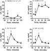
LH responses (mean ± SEM) in catheterized GD85 lamb fetuses given IV bolus injections of (A) saline (S, 500 µL), (B) GnRH (G; 25 µg), (C,D) murine kisspeptin-10 (K, 10 µg). Acute experiments were performed while the animals were under general anesthesia. Control vehicle and drugs were injected into the jugular vein, and blood samples obtained from the carotid artery before (–15 and 0 minutes) or at 30, 60, 90, and 120 minutes after injection. Data were analyzed by 1-way ANOVA for repeated measures followed by Bonferroni’s multiple comparison tests. *P < .05 vs time = –15 minutes.
Figure 2.
Figure 2.
Testosterone responses (mean ± SEM) to KP-10 infusion in male and female GD85 fetuses. KP-10 stimulated release of LH was accompanied by a rise in serum testosterone concentrations that was offset by 30 minutes. in males but not females. See Fig. 1 for experimental details. Data were analyzed by 1-way ANOVA for repeated measures followed by Bonferroni’s multiple comparison tests. *P < .05 vs time = –15 minutes.
Figure 3.
Figure 3.
LH and testosterone responses to IV KP-10 are abolished 1 hour after pretreatment with the GnRH-receptor antagonist acyline (60 µg/kg, IM). Acyline was injected into neck muscle prior to catheterization. Blood sampling was performed as described in Fig. 1.
Figure 4.
Figure 4.
LH responses (mean ± SEM) in chronically catheterized GD85 lamb fetuses given IV bolus injections of (A) saline (S, 500 µL); (B) mouse kisspeptin-10 (K, 10 µg); Senktide (Sk, 50 µg), and GnRH (G; 50 µg) at time 0. Fetuses were fitted with indwelling jugular vein and carotid artery catheters on GD82 for drug infusion and blood collection, respectively. Chronic experiments were performed on unanesthetized fetuses between GD85 and GD89. Data were analyzed by 2-way ANOVA (sex × time) for repeated measures followed by Bonferroni’s multiple comparison tests. When there was a significant interaction between sex and time, the time effect was evaluated individually for each sex using a 1-way ANOVA for repeated measures. *P < .05 vs time = –30 minutes. †P < .05 male vs female.
Figure 5.
Figure 5.
Digital immunofluorescence images showing colocalization of kisspeptin (Kiss, green fluorescence, FITC) positive perikarya and NKB (red fluorescence, Cy3) in the arcuate nucleus of GD60 and GD85 female lamb fetuses. Note that colocalization of kisspeptin and NKB is shown in the last panel as yellow perikarya. Scale bar = 50 µm.
Figure 6.
Figure 6.
Age and sex comparison of the average (±SEM) number of kisspeptin- (Kiss) and NKB-positive cells in the middle arcuate nucleus of GD60 and GD85 fetal lambs. Also shown are age and sex comparisons for the mean (±SEM) percentage of kisspeptin cells that colocalized NKB and NKB cells that colocalized kisspeptin. Data were analyzed by 2-way (sex × age) ANOVA followed by Tukey’s multiple comparison test. Bars with different letters are significantly different, P < .05.
Figure 7.
Figure 7.
Fluorescent in situ hybridization (FISH) detection of kisspeptin (KISS1), neurokinin B/tachykinin3 (TAC3) and prodynorphin (PDYN) mRNA. (A) Confocal image (X60 magnification) showing representative cells in the ARC of a GD86 fetus expressing kisspeptin (white), NKB (green), and Dyn (red). Section was counterstained using DAPI (blue) for visualization of cell nuclei. (B) Higher magnification view of cell identified in the square in A. Scale bar = 25 μm (A) and 10 μm (B).
Figure 8.
Figure 8.
Sex comparisons of KNDy-related gene expression in the MBH of GD85 fetal lambs. Quantitative real-time polymerase chain reaction was used to measure the gene expression for kisspeptin (KISS1), kisspeptin receptor (KISS1R), NKB (TAC3), NKB receptor (TACR3), prodynorphin (pDYN) and Κ-opioid receptor (OPRK1). Bars represent the fold expression (mean ± SEM) relative to the males. Statistical comparison between sexes were made with Student’s t-test.

Similar articles

Cited by

References

    1. Foster DL, Jackson LM, Padmanabhan V. Novel concepts about normal sexual differentiation of reproductive neuroendocrine function and the developmental origins of female reproductive dysfunction: the sheep model. In: Juengel JL, Murray JF, Smith MF, eds. Reproduction in Domestic Ruminants. Vol 6. Nottingham: Nottingham University Press; 2007:83–107. - PubMed
    1. Clarke IJ, Scaramuzzi RJ, Short RV. Sexual differentiation of the brain: endocrine and behavioural responses of androgenized ewes to oestrogen. J Endocrinol. 1976;71(1):175–176. - PubMed
    1. McCarthy MM, Arnold AP. Reframing sexual differentiation of the brain. Nat Neurosci. 2011;14(6):677–683. - PMC - PubMed
    1. Arnold AP, McCarthy MM. Sexual Differentiation of the Brain and Behavior: A Primer. In: Pfaff DW, Volkow ND, eds. Neuroscience in the 21st Century. New York, NY: Springer New York; 2016:1–30.
    1. Cooke B, Hegstrom CD, Villeneuve LS, Breedlove SM. Sexual differentiation of the vertebrate brain: principles and mechanisms. Front Neuroendocrinol. 1998;19(4):323–362. - PubMed

Publication types

MeSH terms