

Comparative pathology of human and canine myxomatous mitral valve degeneration: 5HT and TGF-β mechanisms
- PMID: 32006823
- PMCID: PMC7078050
- DOI: 10.1016/j.carpath.2019.107196
Comparative pathology of human and canine myxomatous mitral valve degeneration: 5HT and TGF-β mechanisms
Abstract
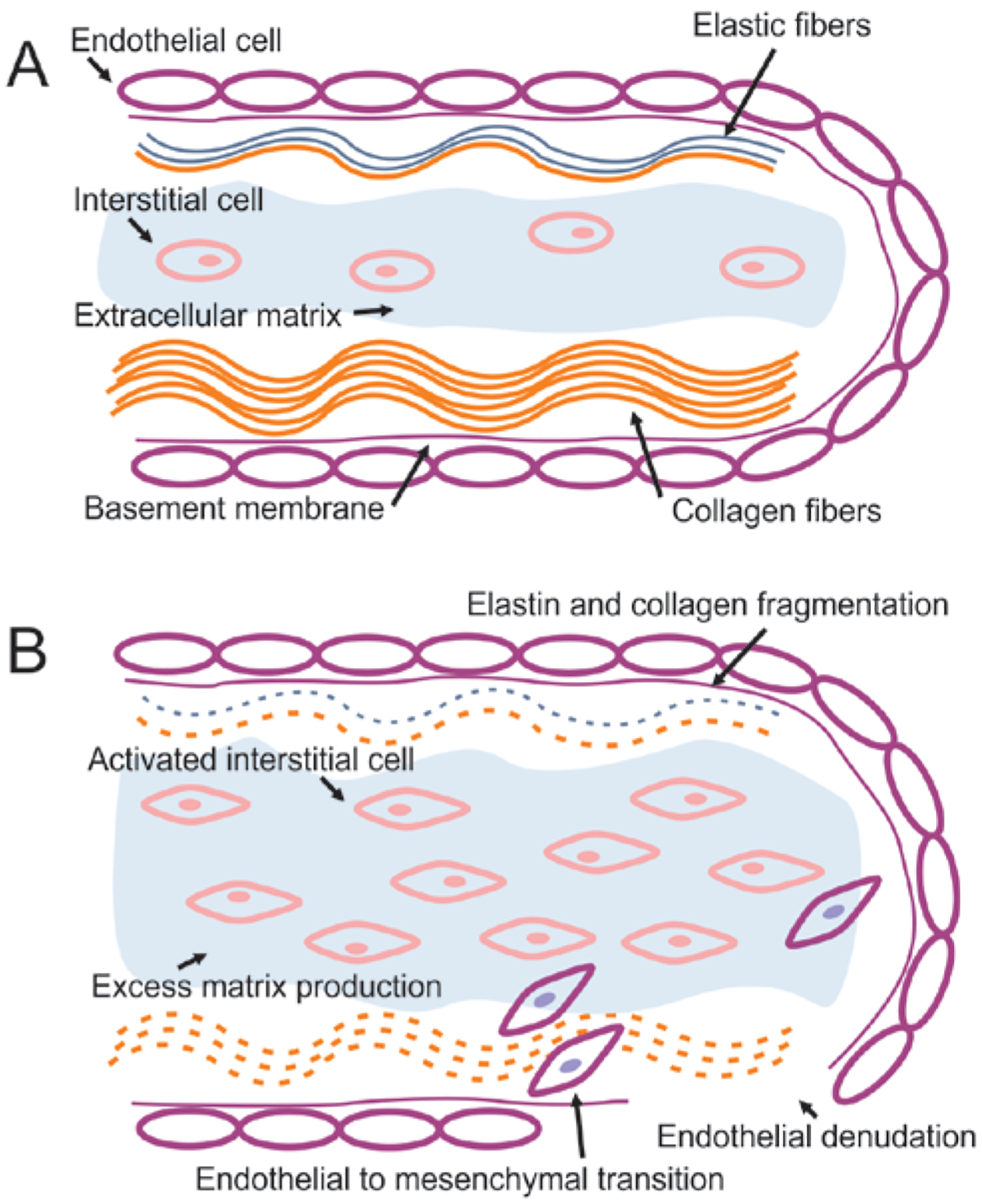
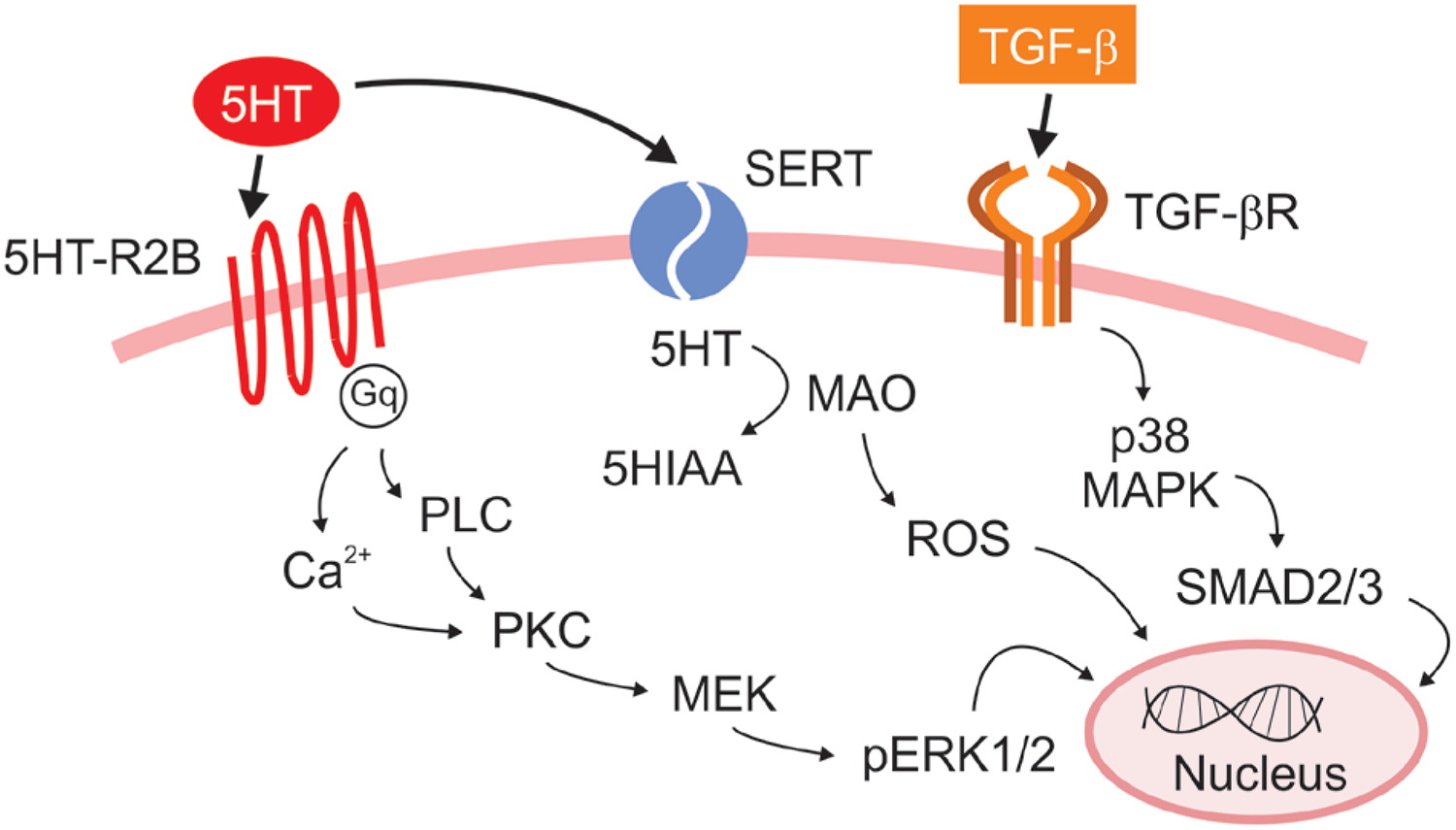
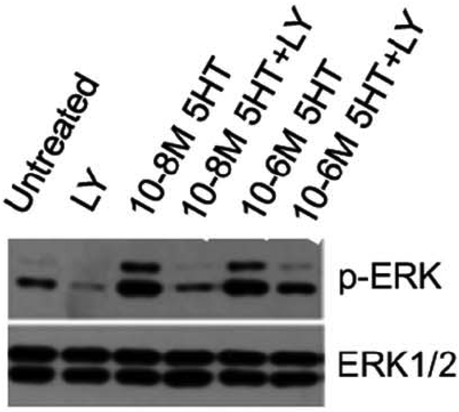
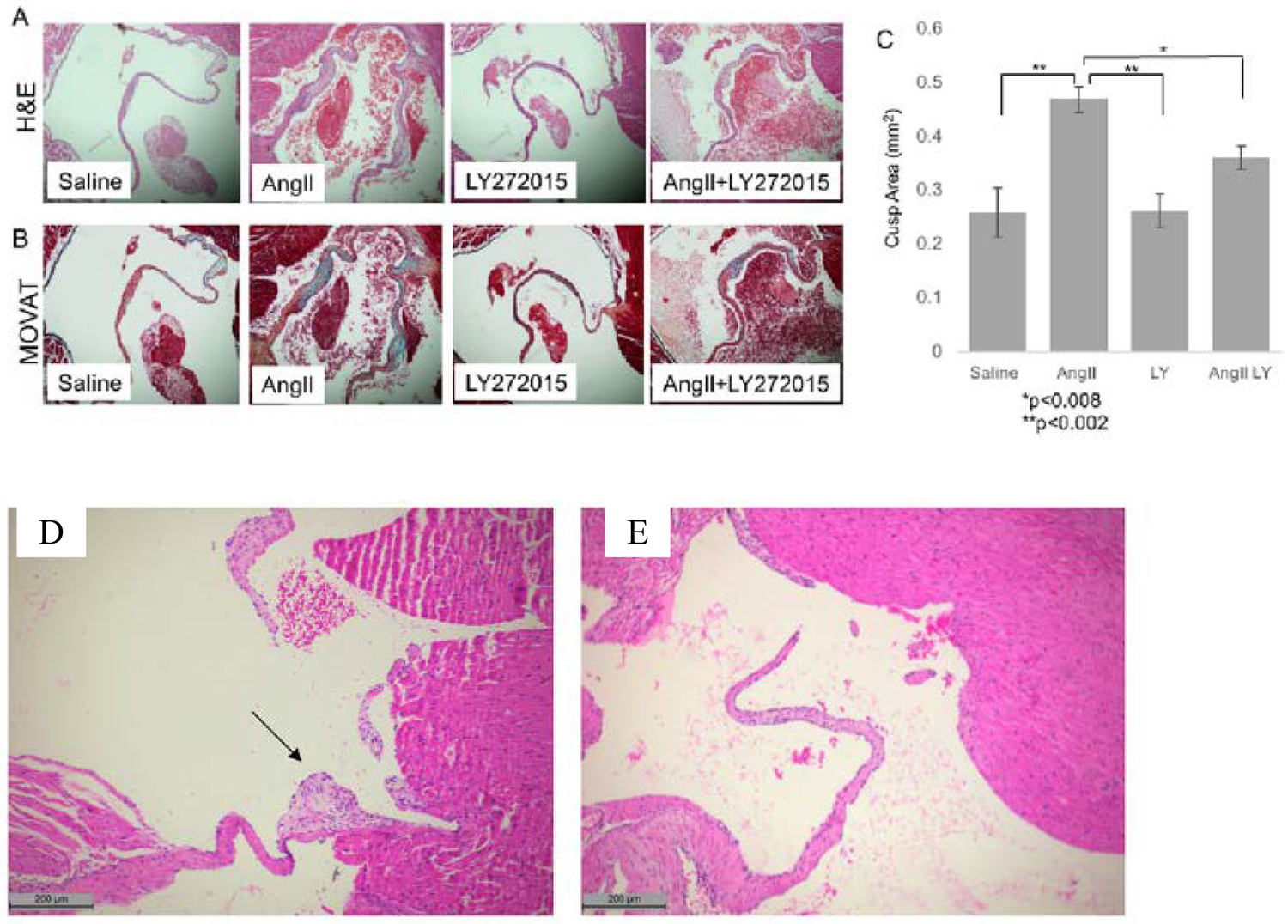
Myxomatous mitral valve degeneration (MMVD) is a leading cause of valve repair or replacement secondary to the production of mitral regurgitation, cardiac enlargement, systolic dysfunction, and heart failure. The pathophysiology of myxomatous mitral valve degeneration is complex and incompletely understood, but key features include activation and transformation of mitral valve (MV) valvular interstitial cells (VICs) into an active phenotype leading to remodeling of the extracellular matrix and compromise of the structural components of the mitral valve leaflets. Uncovering the mechanisms behind these events offers the potential for therapies to prevent, delay, or reverse myxomatous mitral valve degeneration. One such mechanism involves the neurotransmitter serotonin (5HT), which has been linked to development of valvulopathy in a variety of settings, including valvulopathy induced by serotonergic drugs, Serotonin-producing carcinoid tumors, and development of valvulopathy in laboratory animals exposed to high levels of serotonin. Similar to humans, the domestic dog also experiences naturally occurring myxomatous mitral valve degeneration, and in some breeds of dogs, the lifetime prevalence of myxomatous mitral valve degeneration reaches 100%. In dogs, myxomatous mitral valve degeneration has been associated with high serum serotonin, increased expression of serotonin-receptors, autocrine production of serotonin within the mitral valve leaflets, and downregulation of serotonin clearance mechanisms. One pathway closely associated with serotonin involves transforming growth factor beta (TGF-β) and the two pathways share a common ability to activate mitral valve valvular interstitial cells in both humans and dogs. Understanding the role of serotonin and transforming growth factor beta in myxomatous mitral valve degeneration gives rise to potential therapies, such as 5HT receptor (5HT-R) antagonists. The main purposes of this review are to highlight the commonalities between myxomatous mitral valve degeneration in humans and dogs, with specific regards to serotonin and transforming growth factor beta, and to champion the dog as a relevant and particularly valuable model of human disease that can accelerate development of novel therapies.
Keywords: Mitral valve disease; Mitral valve repair; Mitral valve replacement; Myxomatous mitral valve degeneration; Serotonin.
Copyright © 2019. Published by Elsevier Inc.
Conflict of interest statement
Figures
References
-
- Nkomo VT, et al. , Burden of valvular heart diseases: a population-based study. Lancet, 2006. 368(9540): p. 1005–11. - PubMed
-
- Iung B, et al. , A prospective survey of patients with valvular heart disease in Europe: The Euro Heart Survey on Valvular Heart Disease. Eur Heart J, 2003. 24(13): p. 1231–43. - PubMed
-
- Marechaux S, et al. , Functional anatomy and pathophysiologic principles in mitral regurgitation: Non-invasive assessment. Prog Cardiovasc Dis, 2017. 60(3): p. 289–304. - PubMed
-
- Fox PR, Pathology of myxomatous mitral valve disease in the dog. J Vet Cardiol, 2012. 14(1): p. 103–26. - PubMed
Publication types
MeSH terms
Substances
Grants and funding
LinkOut - more resources
Full Text Sources
Medical
