. 2020 May;28(5):669-674.
doi: 10.1016/j.joca.2020.01.011. Epub 2020 Jan 30.

Role of TLR2 and TLR4 in regulation of articular chondrocyte homeostasis

Affiliations

Role of TLR2 and TLR4 in regulation of articular chondrocyte homeostasis

Y Wang et al. Osteoarthritis Cartilage. 2020 May.

Abstract

Objective: Toll-like receptor (TLR)-mediated catabolic responses are implicated to contribute to osteoarthritis (OA). However, deficiency of TLRs has little chondroprotection in mice in vivo. Here, we studied the effect of deficiency of TLR2 and TLR4 in articular chondrocytes on cellular stress responses in vitro.

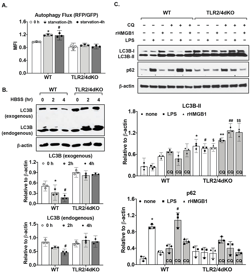
Design: Chondrocytes isolated from TLR2 and TLR4 double knockout (TLR2/4dKO) and wild type (WT) mice and recombinant HMGB1 (rHMGB1) and LPS were used. Expression of anti-oxidant and DNA repair enzymes including SOD1, SOD2 and OGG1, and phosphorylation of H2AX (a marker for DNA damage) were examined by Western blotting. MitoSOX Red staining was used for assessing mitochondrial superoxide generation. Autophagic activity was monitored by flow cytometry analysis of mean fluorescence intensity (MFI) of GFP and RFP in chondrocytes transfected with a tandem GFP-mRFP-LC3 plasmid, and by Western blot analysis of expression of LC3 and p62, a selective autophagy adaptor.

Results: Basal expression of SOD2 but not SOD1 was largely reduced in TLR2/4dKO compared to WT chondrocytes, correlated with significantly enhanced menadione-induced mitochondrial superoxide generation (2.85-3.92 and 3.39 to 8.97 with mean difference 3.39 and 6.18 for 25 and 50μM menadione, respectively) and phosphorylation of H2AX. LPS and rHMGB1 induced expression of SOD2, OGG1 and p62 in WT but not TLR2/4dKO chondrocytes. Autophagy flux was impaired in TLR2/4dKO chondrocytes after acute nutrient stress and by LPS and rHMGB1.

Conclusions: TLR2 and TLR4 deficiency appears to reduce chondrocyte anti-oxidative stress and autophagy flux capacity, which may compromise cartilage homeostasis as a result of chondrocyte dysfunction.

Keywords: Autophagy flux; Chondrocytes; Oxidative stress; Toll-like receptors.


Conflict of interest statement

Competing interests: None

Figures

Figure 1.
Figure 1.. Chondrocytes deficient in TLR2 and TLR4 exhibited reduced anti-oxidative stress capacity.
WT and TLR2/4dKO chondrocytes at basal levels (A) or after stimulation with LPS (1 μg/ml) and rHMGB1 (2 μg/ml) for 18 hours (D) were examined for expression of SOD1, SOD2, OGG1 or Ku70 by Western blotting. β-actin was included as a loading control. WT and TLR2/4dKO chondrocytes were treated with menadione at 25 and 50 μM for 1 hour, mitochondrial superoxide generation (B) was assessed by flow cytometry analysis of mean fluorescent intensity (MFI). Phosphorylation and expression of H2AX were examined by Western blotting (C). Densitometry analysis of Western blot data from 3 independent experiments was included. Student t-test (A) and repeated measures two-way ANOVA analysis (B,C,D) were used for statistical analysis. There was a significant interaction between the effect of menadione and genotype on mitochondrial superoxide generation in B (F(2, 8)=91.73, p<0.0001) and on normalized phosphorylation of H2AX in C (F(2, 8)=9.91, p=0.0068). There was also a significant interaction between the effect of treatment (LPS or rHMGB1) and genotype on normalized SOD2 expression F(2, 8)=79.18, p<0.0001 and normalized OGG1 expression F(2, 8)= 23.46, p=0.0005, but not normalized Ku70 expression. Sidak’s test on the effect of menadione at each concentration between WT and TLR2/4dKO (B, C) and Tukey’s test on the effect of each treatment within WT or TLR2/4dKO (B,C,D) were followed up. In A, *p=0.002, relative to WT. In B, * p=0.0065, # p<0.0001 relative to none in WT, ** p=0.0003 and ## p=0.003 relative to none in TLR2/4dKO. In C, * p=0.012, # p=0.005 relative to none in WT, **, ## p=0.008 relative to none in TLR2/4dKO. In D. * p=0.0004 and # p=0.03 for SOD2, ** p=0.045 and ## p=0.021 for OGG1, relative to none in WT.
Figure 2.
Figure 2.. Autophagy flux was impaired in chondrocytes deficient in TLR2 and TLR4.
Both WT and TLR2/4dKO chondrocytes were transfected with a tandem mRFP and GFP fluorescent-tagged LC3 (ptfLC3) plasmid for 48 hours. The cells were then subjected to nutrient starvation in HBSS for 2 and 4 hours, followed by flow cytometry analysis of mean fluorescence intensity of RFP and GFP. Autophagy flux was determined by the ratio of RFP/GFP (A). Expression of endogenous and exogenous LC3B that was appeared as higher molecular weight conjugates of mRFP-GFP-LC3 on the same blot was examined by Western blotting (B). WT and TLR2/4dKO chondrocytes were stimulated with LPS (1 μg/ml) and rHMGB1 (2 μg/ml) for 18 hours in the presence or absence of choloroquine (CQ, 50 μM). Expression of p62 and LC3B was examined by Western blotting (C). β-actin was included as a loading control. Densitometry analysis of Western blot data from 3 independent experiments was included. Repeated measures two-way ANOVA was used for statistical analysis. There was a significant interaction between the effect nutrient starvation and genotype on autophagy flux (RFP/GFP) F(2, 8)=18.85, p=0.0009) in A, on normalized exogenous LC3 expression F(2, 8)=33.18, p=0.0001 and normalized endogenous LC3 expression F(2, 8)=16.7, p=0.001) in B. There was also a significant interaction between treatment (LPS or rHMGB1) and genotype on normalized LC3B-II expression F(5, 20)=11.46, p<0.0001 and normalized p62 expression F(5, 20)=48, p<0.0001 in C. Tukey’s test on the effect of nutrient starvation or treatment within WT or TLR2/4dKO (A,B,C) and Sidak’s test on the effect of each treatment between WT and TLR2/4dKO (C) were followed up. In A, *p=0.007, #p=0.049, relative to 0 h. In B, * p=0.04, # p=0.005 relative to 0 hour in WT for exogenous LC3B, #p=0.017 relative to 0 hour in WT for endogenous LC3B. For LC3B-II in C, * p=0.0009, # p=0.0002, $ p=0.014 relative to none, LPS and rHMGB1, respectively, in WT, ** p=0.003, ## p=0.001, $$ p=0.018 relative to none, LPS and rHMGB1 respectively, in WT in the presence of choloroquine. For p62 in C, * p=0.01 and # p=0.018 relative to none in WT and WT in the presence of choloroquine, respectively.

Similar articles

Cited by

References

    1. Loeser RF, Goldring SR, Scanzello CR, Goldring MB. Osteoarthritis: a disease of the joint as an organ. Arthritis Rheum. 2012;64:1697–707. - PMC - PubMed
    1. Liu-Bryan R. Synovium and the innate inflammatory network in osteoarthritis progression. Curr Rheumatol Rep. 2013;15:323. - PMC - PubMed
    1. Blom AB, van Lent PL, Abdollahi-Roodsaz S, van der Kraan P, van den Berg W. Elusive role for toll like receptor 2 in joint pathology during experimental osteoarthritis. Osteoarthritis and Cartilage. 2011;19(Suppl 1):25.
    1. Nasi S, Ea HK, Chobaz V, van Lent P, Lioté F, So A, et al. Dispensable role of myeloid differentiation primary response gene 88 (MyD88) and MyD88-dependent toll-like receptors (TLRs) in a murine model of osteoarthritis. Joint Bone Spine. 2014;81:320–4. - PubMed
    1. Kalaitzoglou E, Lopes EBP, Fu Y, Herron JC, Flaming JM, Donovan EL, Hu Y, Filiberti A, Griffin TM, Humphrey MB. TLR4 Promotes and DAP12 Limits Obesity-Induced Osteoarthritis in Aged Female Mice. JBMR Plus. 2018;3:e10079. - PMC - PubMed

Publication types

MeSH terms