. 2020 Dec;77(24):5207-5221.
doi: 10.1007/s00018-020-03465-3. Epub 2020 Feb 1.

Thalidomide targets EGFL6 to inhibit EGFL6/PAX6 axis-driven angiogenesis in small bowel vascular malformation

Affiliations

Thalidomide targets EGFL6 to inhibit EGFL6/PAX6 axis-driven angiogenesis in small bowel vascular malformation

Chao-Tao Tang et al. Cell Mol Life Sci. 2020 Dec.

Abstract

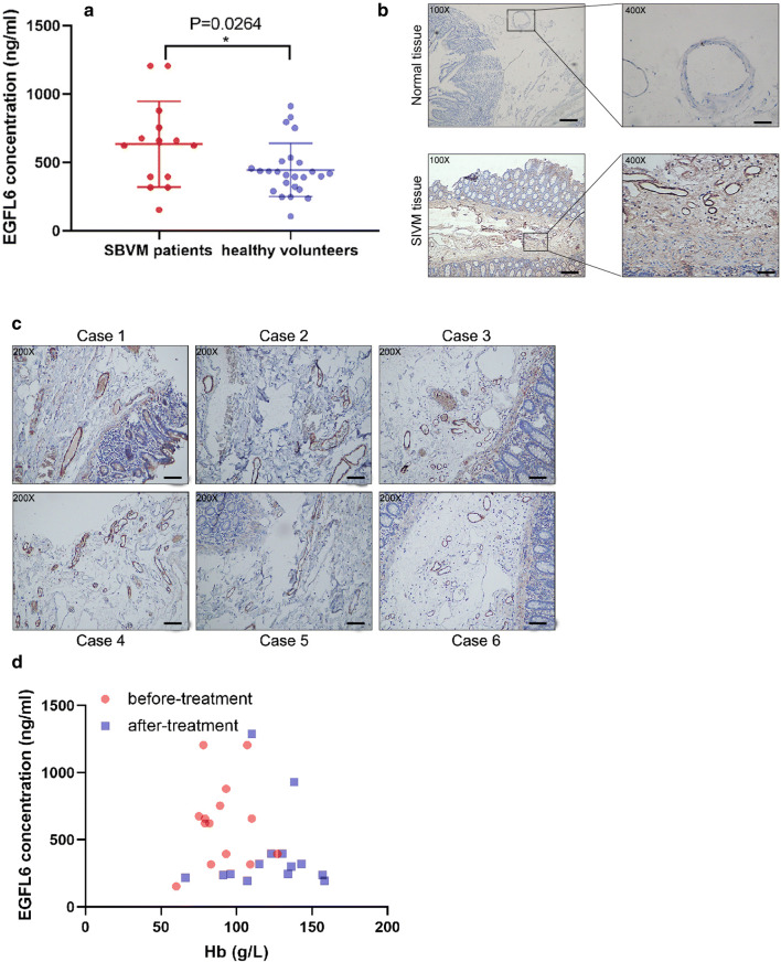
Background: Small bowel vascular malformation disease (SBVM) is the most common cause of obscure gastrointestinal bleeding (OGIB). Several studies suggested that EGFL6 was able to promote the growth of tumor endothelial cells by forming tumor vessels. To date, it remains unclear how EGFL6 promotes pathological angiogenesis in SBVM and whether EGFL6 is a target of thalidomide.

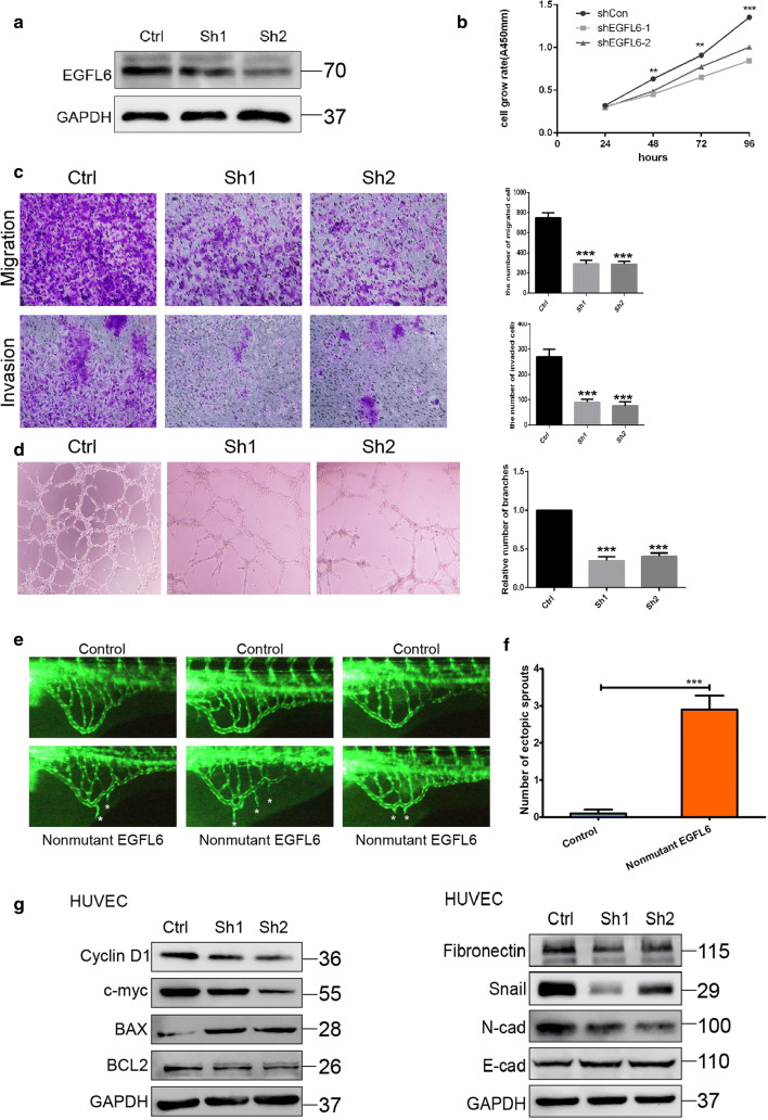
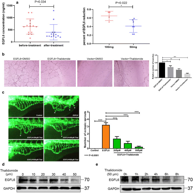
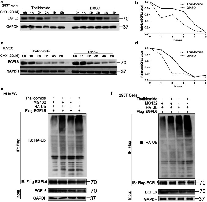
Methods: We took advantage of SBVM plasma and tissue samples and compared the expression of EGFL6 between SBVM patients and healthy people via ELISA and Immunohistochemistry. We elucidated the underlying function of EGFL6 in SBVM in vitro and by generating a zebrafish model that overexpresses EGFL6, The cycloheximide (CHX)-chase experiment and CoIP assays were conducted to demonstrate that thalidomide can promote the degradation of EGFL6 by targeting CRBN.

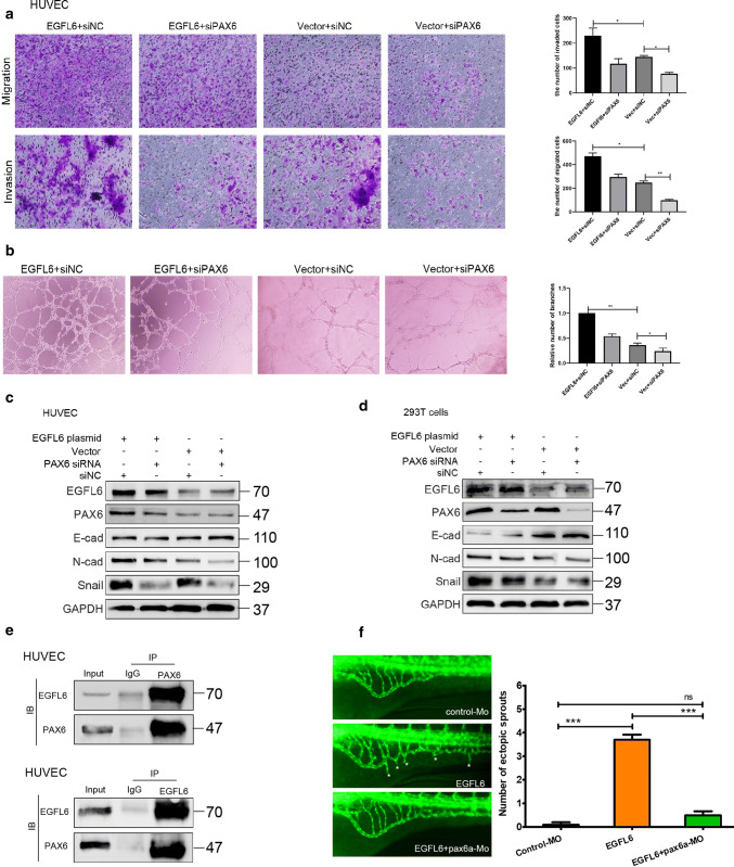
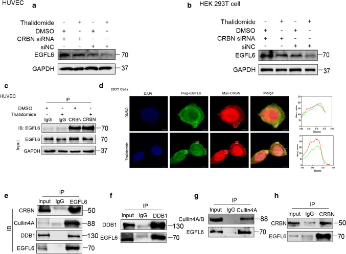
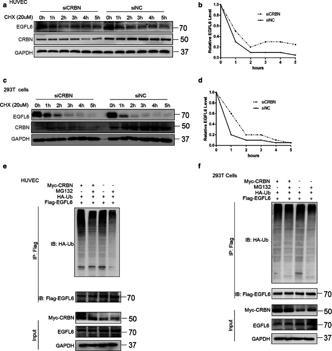
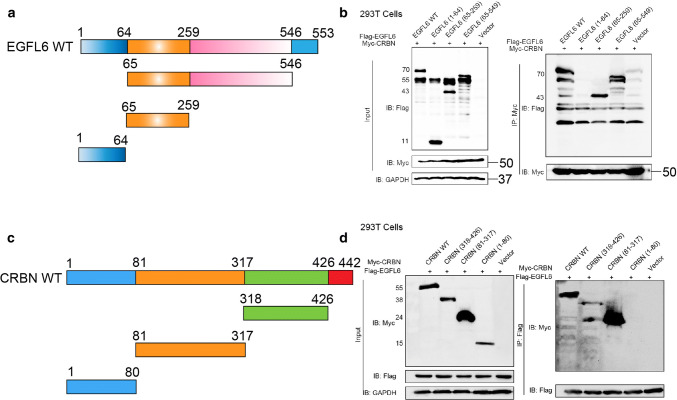
Results: The analysis of SBVM plasma and tissue samples revealed that EGFL6 was overexpressed in the patients compared to healthy people. Using in vitro and in vivo assays, we demonstrated that an EMT pathway triggered by the EGFL6/PAX6 axis is involved in the pathogenesis of SBVM. Furthermore, through in vitro and in vivo assays, we elucidated that thalidomide can function as anti-angiogenesis medicine through the regulation of EGFL6 in a proteasome-dependent manner. Finally, we found that CRBN can mediate the effect of thalidomide on EGFL6 expression and that the CRBN protein interacts with EGFL6 via a Lon N-terminal peptide.

Conclusion: Our findings revealed a key role for EGFL6 in SBVM pathogenesis and provided a mechanism explaining why thalidomide can cure small bowel bleeding resulting from SBVM.

Keywords: Angiodysplasia; Obscure gastrointestinal bleeding; Proteasome-dependent degradation.


Conflict of interest statement

The authors disclose no conflicts.

Figures

Fig. 1
Fig. 1
EGFL6 expression is upregulated in SBVM tissues and serum of patients and correlates with the level of Hb. a ELISA was used to determine the level of EGFL6 in plasma between healthy volunteers and SBVM patients. b Representative images of IHC staining of EGFL6 in SBVM and normal tissues. Scale bars: left, 200 µm; right, 50 µm. c EGFL6 protein expressions in six SBVM samples were analyzed by IHC. Scale bars: 100 µm. d Correlation between EGFL6 expression and hemoglobin (Hb) level was analyzed by Graphpad software
Fig. 2
Fig. 2
EGFL6 knockdown inhibits proliferation and angiogenesis in vivo and in vitro. a Western blot assay was used to determine the effect of EGFL6 siRNA. b cell viability was measured by CCK8 assay with HUVEC transfected with siNC and EGFL6 siRNA, Data are shown as the mean ± SEM. c cells migration and invasion assay with HUVEC transfected with siNC and EGFL6 siRNA, represented image was shown on the left and mean ± SEM was shown by bar chart. d Tuber formation assay with HUVEC transfected with siNC and EGFL6 siRNA, represented image was shown on the left and Mean ± SEM was shown by bar chart, *P < 0.05, **P < 0.01. e Representative fluorescent images of uninjected control or embryos injected with 100 pg nonmutant human EGFL6 mRNA at 3,dpf. The subintestinal vessels form on the dorsolateral surface of the yolk on both sides of the embryo in the shape of a basket over the yolk. Compared with control, higher magnification of the SIV revealed that ectopic branches (asterisk) can be observed after nonmutant human EGFL6 mRNA injection, dpf, days postfertilization; SIV, subintestinal vein. f Quantification of the average number of ectopic SIV segments. ***P < 0.001 (n = 10; Student’s t test). g The expressions of molecules related with cell proliferation (left) and cell migration (right) were detected with western blot. Data represent the average of three independent experiments
Fig. 3
Fig. 3
PAX6 is involved in EGFL6-mediated angiogenesis in vitro and in vivo. a cells’ migration and invasion assay with HUVEC that were treated with EGFL6 plasmid combined with PAX6 siRNA; represented image was shown on the left and Quantitative data are expressed as mean ± SEM, **P < 0.01, ***P < 0.001. b Tuber formation assay with HUVEC that were treated with EGFL6 plasmid combined with PAX6 siRNA; represented image was shown on the left and result was shown by bar chart, **P < 0.01, ***P < 0.001. c, d The expression of molecules related with cell migration in HUVEC and HEK293T cells that were treated with EGFL6 plasmid combined with PAX6 siRNA were detected with western blot. e Interaction between endogenous PAX6 and EGFL6 was detected by co-immunoprecipitation assay with anti-EGFL6 and anti-PAX6 antibody in HUVEC. The immunoglobulin G (IgG) antibody bought from BioSharp company was used as the control group and cell lysates were exploited to examine the expression of EGFL6 and PAX6. f Representative fluorescent images of zebrafish embryos at 72 hpf treated with control-MO (4 ng per embryo), EGFL6 (100 pg per embryo), or EGFL6 plus pax6a-MO (4 ng per embryo). Compared with EGFL6 overexpression embryos, embryos co-administered with pax6a-MO and EGFL6 present a decreased number of ectopic SIV segments (asterisk), quantification of the average number of ectopic SIV segments at 72 hpf. (n = 10; ANOVA; ***P < 0.0001; ns not significant.). Data represent the average of three independent experiments
Fig. 4
Fig. 4
Thalidomide inhibits angiogenesis by targeting EGFL6 in vitro and in vivo. a ELISA assay was performed to detect the expression of EGFL6 in 14 SBVM patients who were treated with thalidomide; picture on the left was the alteration of EGFL6 expression between before treatment and after treatment; picture on the right was the percent of EGFL6 reduction between patients treated with 100 mg per day and patients treated with 50 mg per day. b Tuber formation assay with HUVEC that were treated with EGFL6 plasmid combined with 50 μM thalidomide; represented image was shown on the left; and result was shown by bar chart, **P < 0.01, ***P < 0.001. c Representative fluorescent images of zebrafish embryos at 72 hpf treated with 0.1% dimethyl sulfoxide (DMSO) (control) and EGFL6 (200 pg per embryo) or co-administered with thalidomide (200 μM, 400 μM, 800 μM) for 24 h. Compared with EGFL6 overexpression embryos, embryos’ treatment with thalidomide present a decreased number of ectopic SIV segments (asterisk), quantification of the average number of ectopic SIV segments at 72 hpf. n = 10; ANOVA; ***P < 0.0001. d HUVEC were treated with thalidomide at different concentration and western blot assay was performed to determine the expression of EGFL6. e HUVEC were treated with 50 μm thalidomide at different time point and cell lysate were subjected to western blot assay to determine the expression of EGFL6
Fig. 5
Fig. 5
Thalidomide degrades EGFL6 by enhancing its ubiquitination level. a, d HEK293T cells and HUVEC were treated with 50 μm thalidomide combined with 20 μM CHX at indicated time. a HEK293T cells lysate were subjected to western blot assay to determine the expression of EGFL6. b The immunoblotting of EGFL6 was quantified by Image J. c HUVEC lysate were subjected to western blot assay to determine the expression of EGFL6. d The immunoblotting of EGFL6 was quantified by Image J. e, f HA-Ub and Flag-EGFL6 plasmids were co-transfected in HEK293T cells. After 48 h, cells were treated with 20 μm MG132 and 50 μm for 6 h. EGFL6 ubiquitination level was assessed by immunoprecipitation and EGFL6 expression after treatment of thalidomide and MG132 was detected by western blot
Fig. 6
Fig. 6
CRBN regulates the degradation of EGFL6 induced by thalidomide. a, b HEK293T cells and HUVEC were treated with CRBN siRNA combined with 50 μM thalidomide; western blot was performed to determine the expression of EGFL6. c HUVEC were treated with 50 μM thalidomide. Co-immunoprecipitataion assay was performed to explore the interaction between EGFL6 and CRBN. d HUVEC were transfected with Flag-EGFL6 plasmid and Myc-CRBN plasmid, after 48 h, cells were treated with thalidomide. Immunofluorescence assay was performed to detect the localization between EGFL6 and CRBN. e, h Co-immunoprecipitataion assay was performed to determine the interaction between EGFL6 and CRBN–DDB1–Cullin 4A complex
Fig. 7
Fig. 7
CRBN degrades EGFL6 by enhancing its ubiquitination level. a, d HEK293T cells and HUVEC were treated with CRBN siRNA combined with 20 μM CHX at indicated time. a HUVEC lysate were subjected to western blot assay to determine the expression of EGFL6. b The immunoblotting of EGFL6 was quantified by Image J. c HEK293 cells lysate were subjected to western blot assay to determine the expression of EGFL6. d The immunoblotting of EGFL6 was quantified by Image J. e, f HA-Ub plasmid, Flag-EGFL6 plasmid and CRBN siRNA were co-transfected in HEK293T cells. After 48 h, cells were treated with 20 μm MG132 for 6 h. EGFL6 ubiquitination level was assessed by immunoprecipitation and EGFL6 expression was detected by western blot
Fig. 8
Fig. 8
CRBN interacts with EGFL6. a A schematic diagram shows the structural domains of EGFL6 according to Uniprot website. b The control vector, EGFL6 wide-type (WT) plasmid, and truncated mutants of EGFL6 were co-transfected with Myc,CRBN WT plasmid into HEK293T cells. Co-immunoprecipitataion assay was performed to detect the interaction between EGFL6 and CRBN protein. c A schematic diagram shows the structural domains of CRBN according to Uniprot website. d The control vector, CRBN WT plasmid, and truncated mutants of CRBN were co-transfected with Flag,EGFL6 WT plasmid into HEK293T cells. Co-immunoprecipitataion assay was performed to detect the interaction between EGFL6 and CRBN proteins

Similar articles

Cited by

References

    1. Jackson CS, Strong R. Gastrointestinal angiodysplasia: diagnosis and management. Gastrointest Endosc Clin N Am. 2017;27(1):51–62. doi: 10.1016/j.giec.2016.08.012. - DOI - PubMed
    1. Becq A, et al. Hemorrhagic angiodysplasia of the digestive tract: pathogenesis, diagnosis, and management. Gastrointest Endosc. 2017;86(5):792–806. doi: 10.1016/j.gie.2017.05.018. - DOI - PubMed
    1. Feng N, et al. HIF-1alpha and HIF-2alpha induced angiogenesis in gastrointestinal vascular malformation and reversed by thalidomide. Sci Rep. 2016;6:27280. doi: 10.1038/srep27280. - DOI - PMC - PubMed
    1. Holleran G, et al. Expression of angiogenic factors in patients with sporadic small bowel angiodysplasia. J Clin Gastroenterol. 2015;49(10):831–836. doi: 10.1097/MCG.0000000000000260. - DOI - PubMed
    1. Ge ZZ, et al. Efficacy of thalidomide for refractory gastrointestinal bleeding from vascular malformation. Gastroenterology. 2011;141(5):1629-37.e1-4. doi: 10.1053/j.gastro.2011.07.018. - DOI - PubMed

MeSH terms

LinkOut - more resources