

Optimal Plane Selection for Measuring Post-prandial Blood Flow Increase within the Superior Mesenteric Artery: Analysis Using 4D Flow and Computational Fluid Dynamics
- PMID: 32009062
- PMCID: PMC7809144
- DOI: 10.2463/mrms.mp.2019-0089
Optimal Plane Selection for Measuring Post-prandial Blood Flow Increase within the Superior Mesenteric Artery: Analysis Using 4D Flow and Computational Fluid Dynamics
Abstract
Purpose: 2D cine phase contrast (PC)-MRI is a standard velocimetry for the superior mesenteric artery (SMA); however, the optimal localization of the measurement plane has never been fully discussed previously. The purpose of this Institutional Review Board approved prospective and single arm study is to test whether flow velocimetry of the SMA with combined use of 2D cine PC-MRI and meal challenge is dependent on the localizations of the measurement planes and to seek optimal section for velocimetry.
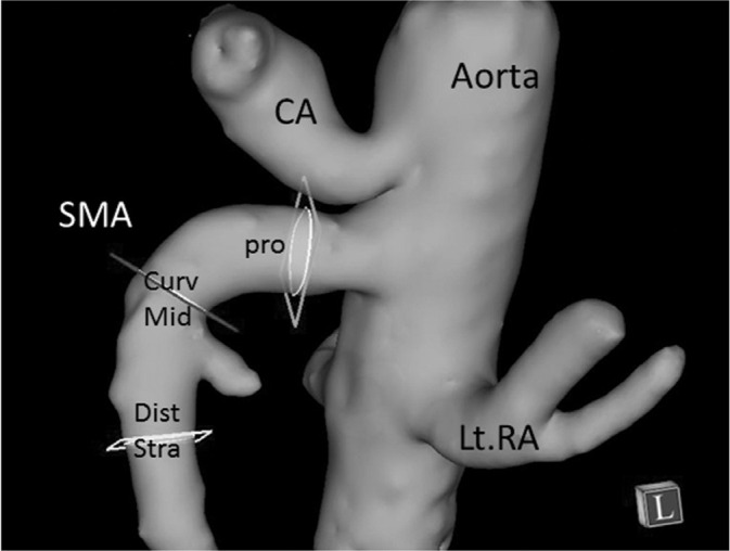
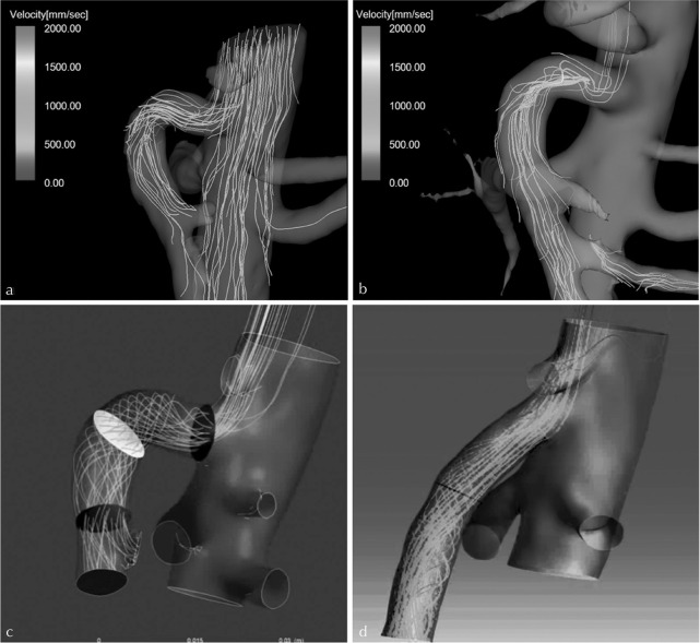
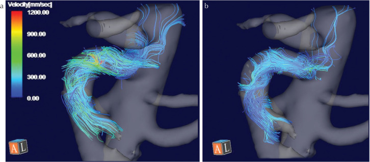
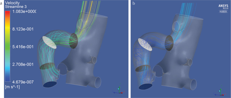
Methods: Seven healthy volunteers underwent cardiac phase resolved ECG gated 2D cine PC-MRI pre- and 30 min post-meal challenge at three measurement planes: proximal, curved mid section and distal straight section of the SMA at 3T. 4D Flow using 3D cine PC-MRI with vastly undersampled isotropic projection imaging (PC VIPR) was also performed right after 2D cine PC-MRI to delineate the flow dynamics within the SMA using streamline analysis. Two radiologists measured flow velocities, and rated the appearances of the abnormal flow in the SMA on streamlines derived from the 4D Flow and the computational fluid dynamics (CFD).
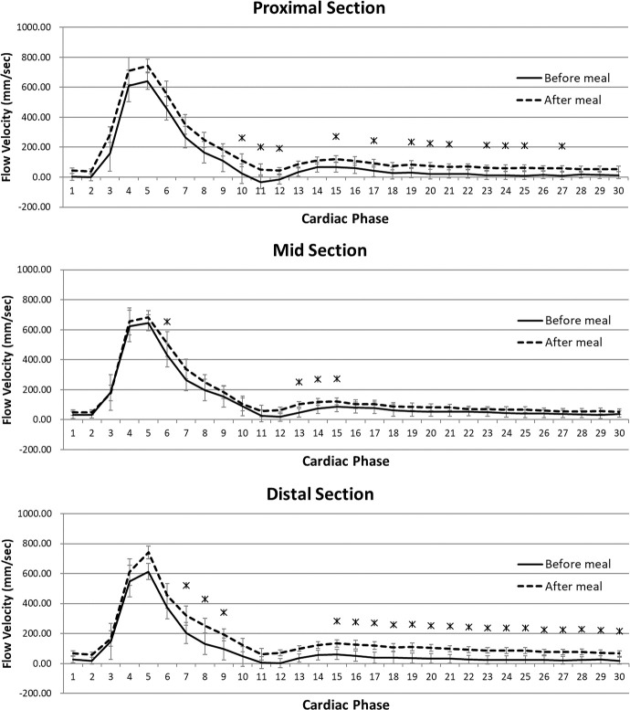
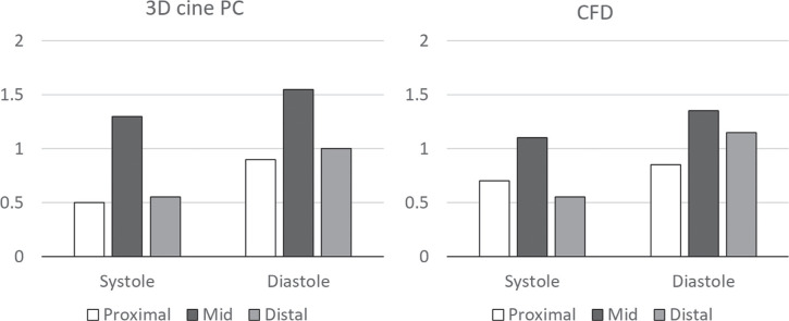
Results: 2D cine PC-MRI measured increased temporally averaged flow velocity (mm/s) after the meal challenge only in the proximal (129.3 vs. 97.8, P = 0.0313) and distal section (166.9 vs. 96.2, P = 0.0313), not in the curved mid section (113.1 vs. 85.5, P = 0.0625). The average velocities were highest and their standard errors (8.5-26.5) were smallest at the distal straight section both before and after the meal challenge as compared with other sections. The streamline analysis depicted more frequent appearances of vertical or helical flow in the curved mid section both on 4D Flow and CFD (κ: 0.27-0.68).
Conclusion: SMA velocimetry with 2D cine PC-MRI was dependent on the localization of the measurement planes. Distal straight section, not in the curved mid section is recommended for MR velocimetry.
Keywords: 4D Flow; flow measurement; phase contrast image; post-prandial hyperemia; superior mesenteric artery.
Conflict of interest statement
The corresponding author Yasuo Takehara is an endowed chair of Nagoya University supported by a private company; however, the status is irrelevant to the contents of the paper.
Coauthors Tetsuya Wakayama, Atsushi Nozaki and Hiroyuki Kabasawa are employees of GE Healthcare.
Other coauthors have nothing to disclose related to the current study.
Figures
References
-
- Debatin JF. Quantification of blood flow, In: Arlart IP, Bongartz GM, Marchal G, eds. Magnetic Resonance Angiography. 2nd ed New York: Springer, 2002.
-
- Li KC, Whitney WS, McDonnell CH, et al. Chronic mesenteric ischemia: evaluation with phase-contrast cine MR imaging. Radiology 1994; 190:175–179. - PubMed
-
- Masui T, Isoda H, Mochizuki T, et al. Effects of meal intake on the flow velocity in the superior mesenteric artery: evaluation with 2D phase mapping MRI. J Comput Assist Tomogr 1994; 18:590–595. - PubMed
-
- Naganawa S, Cooper TG, Jenner G, Potchen EJ, Ishigaki T. Flow velocity and volume measurement of superior and inferior mesenteric artery with cine phase contrast magnetic resonance imaging. Radiat Med 1994; 12:213–220. - PubMed
MeSH terms
LinkOut - more resources
Full Text Sources
Miscellaneous