Review
. 2020 May;380(2):379-392.
doi: 10.1007/s00441-020-03172-2. Epub 2020 Feb 3.

From bedside to bench: lung ultrasound for the assessment of pulmonary edema in animal models

Affiliations
Review

From bedside to bench: lung ultrasound for the assessment of pulmonary edema in animal models

Jana Grune et al. Cell Tissue Res. 2020 May.

Abstract

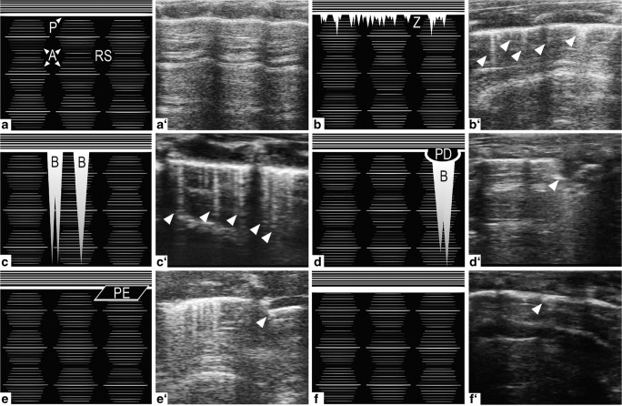
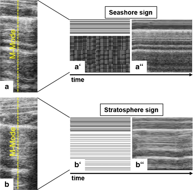
Traditionally, the lung has been excluded from the ultrasound organ repertoire and, hence, the application of lung ultrasound (LUS) was largely limited to a few enthusiastic clinicians. Yet, in the last decades, the recognition of the previously untapped diagnostic potential of LUS in intensive care medicine has fueled its widespread use as a rapid, non-invasive and radiation-free bedside approach with excellent diagnostic accuracy for many of the most common causes of acute respiratory failure, e.g., cardiogenic pulmonary edema, pneumonia, pleural effusion and pneumothorax. Its increased clinical use has also incited attention for the potential usefulness of LUS in preclinical studies with small animal models mimicking lung congestion and pulmonary edema formation. Application of LUS to small animal models of pulmonary edema may save time, is cost-effective, and may reduce the number of experimental animals due to the possibility of serial evaluations in the same animal as compared with traditional end-point measurements. This review provides an overview of the emerging field of LUS with a specific focus on its application in animal models and highlights future perspectives for LUS in preclinical research.

Keywords: Animal models; Diagnostics; Lung ultrasound; Pulmonary edema.


Conflict of interest statement

The authors declare that they have no conflict of interest.

Figures

Fig. 1
Fig. 1
Physiologic and pathologic patterns in LUS B-Mode imaging. (a) Normally aerated lung with a distinct hyperechoic A-line (A) pattern subpleurally and regularly spaced rib shadows (RS). (b) Mostly aerated lung with small, hyperechoic, comet-tail artifacts arising downwards from the pleura (P), the Z-lines (Z). The association of Z-lines with disease states is presently unclear. (c) Partially aerated lung with long, A-line erasing, hyperechoic comet-tail artifacts called B-lines (B) arising downwards from the pleura indicating in this case the presence of alveolar-interstitial syndrome. (d) Pleural defect, resulting in a well-defined interruption of the pleural line. Occasional B-lines arising from the lower edge of the hypoechoic, defective area. (e) Hypoechoic pleural effusion (PE) between parietal (upper) and visceral (lower) pleura lines, which are otherwise indistinguishable by LUS imaging; commonly associated with conditions like heart failure (transudate) or pulmonary embolism (exudate). Also note the presence of Z- and B-lines on the left side of the LUS image. (f) Pleural thickening, indicating the presence of fibrotic or inflammatory lesions
Fig. 2
Fig. 2
M-Mode imaging across supra- and subpleural spaces over time in LUS. (a) Seashore sign: LUS presents suprapleurally with continuous wave-like lines and a diffuse, sand-like pattern subpleurally, indicating physiologic movement of lung tissue during respiration. (b) Stratosphere sign: lung movement is absent, suggesting the occurrence of a pneumothorax

References

    1. Abdalla W, Elgendy M, Abdelaziz AA, Ammar MA. Lung ultrasound versus chest radiography for the diagnosis of pneumothorax in critically ill patients: a prospective, single-blind study. Saudi J Anaesth. 2016;10:265–269. doi: 10.4103/1658-354X.174906. - DOI - PMC - PubMed
    1. Abu-Zidan FM, Hefny AF, Corr P. Clinical ultrasound physics. J Emerg Trauma Shock. 2011;4:501–503. doi: 10.4103/0974-2700.86646. - DOI - PMC - PubMed
    1. Agricola E, Bove T, Oppizzi M, et al. “Ultrasound comet-tail images”: a marker of pulmonary edema: a comparative study with wedge pressure and extravascular lung water. Chest. 2005;127:1690–1695. doi: 10.1378/chest.127.5.1690. - DOI - PubMed
    1. Angus DC, Linde-Zwirble WT, Lidicker J, et al. Epidemiology of severe sepsis in the United States: analysis of incidence, outcome, and associated costs of care. Crit Care Med. 2001;29:1303–1310. doi: 10.1097/00003246-200107000-00002. - DOI - PubMed
    1. Aronchick J, Epstein D, Gefter WB, Miller WT. Evaluation of the chest radiograph in the emergency department patient. Emerg Med Clin North Am. 1985;3:491–505. - PubMed

LinkOut - more resources