Comparative Study
. 2020 Feb 4;9(3):e014035.
doi: 10.1161/JAHA.119.014035. Epub 2020 Feb 3.

Moderate Hypothermia Modifies Coronary Hemodynamics and Endothelium-Dependent Vasodilation in a Porcine Model of Temperature Management

Affiliations
Comparative Study

Moderate Hypothermia Modifies Coronary Hemodynamics and Endothelium-Dependent Vasodilation in a Porcine Model of Temperature Management

Joaquim Bobi et al. J Am Heart Assoc. .

Abstract

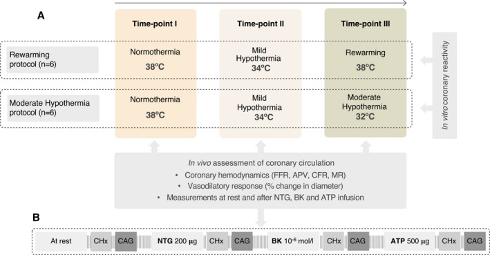
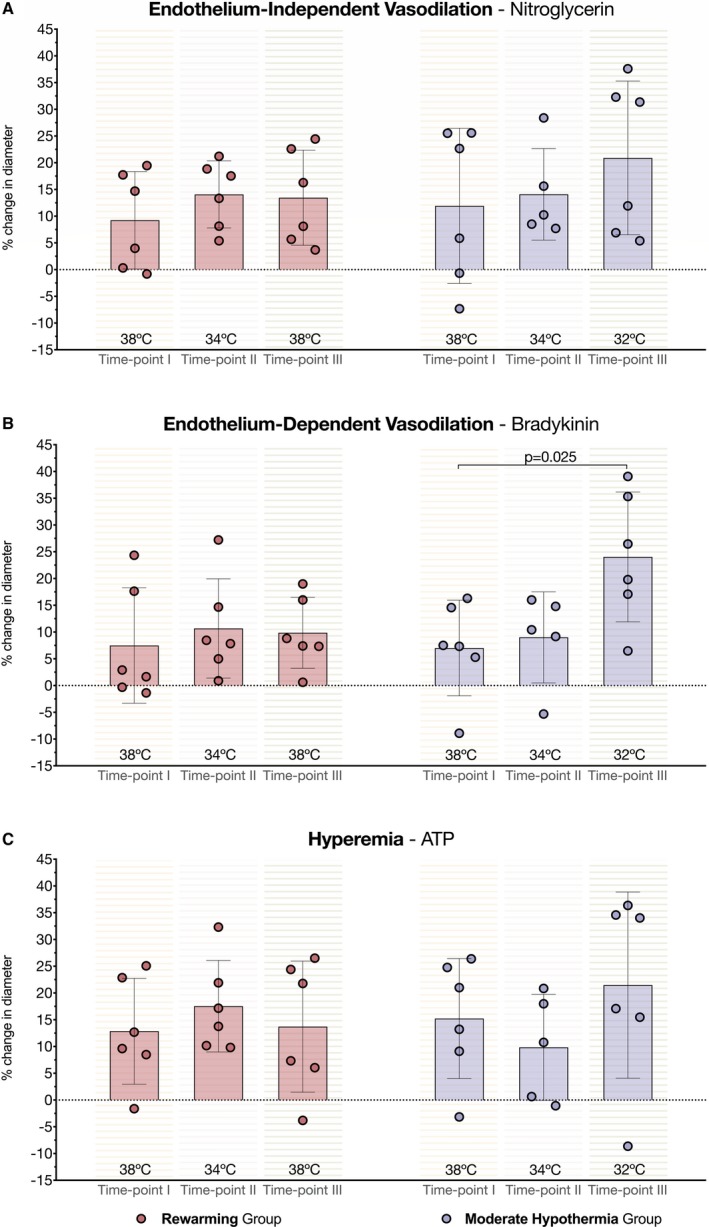
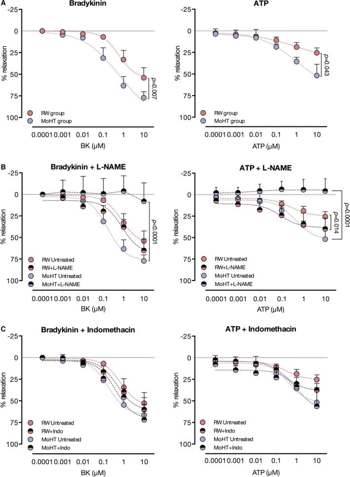
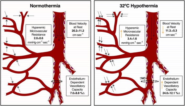
Background Hypothermia has been associated with therapeutic benefits including reduced mortality and better neurologic outcomes in survivors of cardiac arrest. However, undesirable side effects have been reported in patients undergoing coronary interventions. Using a large animal model of temperature management, we aimed to describe how temperature interferes with the coronary vasculature. Methods and Results Coronary hemodynamics and endothelial function were studied in 12 pigs at various core temperatures. Left circumflex coronary artery was challenged with intracoronary nitroglycerin, bradykinin, and adenosine at normothermia (38°C) and mild hypothermia (34°C), followed by either rewarming (38°C; n=6) or moderate hypothermia (MoHT; 32°C, n=6). Invasive coronary hemodynamics by Doppler wire revealed a slower coronary blood velocity at 32°C in the MoHT protocol (normothermia 20.2±11.2 cm/s versus mild hypothermia 18.7±4.3 cm/s versus MoHT 11.3±5.3 cm/s, P=0.007). MoHT time point was also associated with high values of hyperemic microvascular resistance (>3 mm Hg/cm per second) (normothermia 2.0±0.6 mm Hg/cm per second versus mild hypothermia 2.0±0.8 mm Hg/cm per second versus MoHT 3.4±1.6 mm Hg/cm per second, P=0.273). Assessment of coronary vasodilation by quantitative coronary analysis showed increased endothelium-dependent (bradykinin) vasodilation at 32°C when compared with normothermia (normothermia 6.96% change versus mild hypothermia 9.01% change versus MoHT 25.42% change, P=0.044). Results from coronary reactivity in vitro were in agreement with angiography data and established that endothelium-dependent relaxation in MoHT completely relies on NO production. Conclusions In this porcine model of temperature management, 34°C hypothermia and rewarming (38°C) did not affect coronary hemodynamics or endothelial function. However, 32°C hypothermia altered coronary vasculature physiology by slowing coronary blood flow, increasing microvascular resistance, and exacerbating endothelium-dependent vasodilatory response.

Keywords: NO; coronary physiology; endothelium; microcirculation; therapeutic hypothermia.


Figures

Figure 1
Figure 1
Flow chart illustrating study design with the assessments performed at each temperature time point (A) and the protocol of intracoronary drug administration (B). APV indicates average peak velocity; CAG, coronary angiography; CFR, coronary flow reserve; CHx, coronary hemodynamics; FFR fractional flow reserve; MR, microvascular resistance.
Figure 2
Figure 2
Invasive assessment of coronary hemodynamics by Doppler wire. A, Depiction of each coronary hemodynamic variable studied by Doppler wire (black arrow) and its relevance based on coronary vascular compartments. B, Representative coronary angiography showing the retrograde positioning of the Doppler wire in the left circumflex coronary artery (yellow arrow) and a selected branch used to study drug‐induced vasodilation by measuring the diameter before and after each drug challenge (yellow dotted lines). bAPV indicates basal average peak velocity; CFR, coronary flow reserve; ∅, diameter; FFR, fractional flow reserve; MR, microvascular resistance; pAPV, peak average peak velocity; QCA, quantitative coronary analysis.
Figure 3
Figure 3
Effects of temperature on coronary vasodilation. A, Left and right parts of the bar chart show similar percent change in diameter after administration of endothelium‐independent vasodilator (nitroglycerin) along rewarming and moderate hypothermia (MoHT) protocols, respectively. B, Right bars (MoHT protocol) show an effect of temperature on endothelium‐dependent vasodilation with a significant increase in the percent change of diameter after administering bradykinin at 32°C time point. Left part of the bar chart (rewarming protocol) shows no differences in coronary vasodilation after bradykinin administration in the rewarming protocol. C, Bar chart depicts no statistical differences in the percent change in diameter at hyperemia (ATP) between time points in either protocol.
Figure 4
Figure 4
In vitro vasorelaxation of temperature preconditioned coronary rings. A, Left chart depicts increased relaxation of arteries from moderate hypothermia (MoHT), in comparison with the rewarming protocol, when challenged with bradykinin, a pure endothelium‐dependent vasodilator. ATP relaxation curves also showed a greater potential to relax in MoHT rings, compared with rewarming, as shown in the right chart. B, Treatment with the nonselective NO inhibitor, N(ω)‐nitro‐L‐arginine methyl ester (lNAME), resulted in a complete suppression of bradykinin‐ (left chart) and ATP‐induced (right chart) relaxation in coronary rings from the MoHT protocol. On the other hand, lNAME did not modify the bradykinin relaxation curve (left chart) and augmented the potential of relaxation induced by ATP (right chart) in rings from the rewarming protocol. C, Indomethacin, a cyclooxygenase inhibitor, did not modify either bradykinin‐ (left chart) or ATP‐induced (right chart) relaxation curves in either protocol.
Figure 5
Figure 5
Main effects of moderate hypothermia (MoHT) in coronary vasculature. Briefly, MoHT (right) was associated with slowed coronary velocity (long arrows inside the coronary artery), augmented microvascular resistance (represented as grey short arrows pointing out small vessels), and increased endothelium‐derived vasodilatory capacity (dotted lines around the schematic representation of a coronary branch) associated with greater NO activity in comparison with reference parameters at normothermia (left).

References

    1. Alzaga AG, Cerdan M, Varon J. Therapeutic hypothermia. Resuscitation. 2006;70:369–380. - PubMed
    1. Meybohm P, Gruenewald M, Albrecht M, Zacharowski KD, Lucius R, Zitta K, Koch A, Tran N, Scholz J, Bein B. Hypothermia and postconditioning after cardiopulmonary resuscitation reduce cardiac dysfunction by modulating inflammation, apoptosis and remodeling. PLoS One. 2009;4:e7588. - PMC - PubMed
    1. Gong P, Hua R, Zhang Y, Zhao H, Tang Z, Mei X, Zhang M, Cui J, Li C. Hypothermia‐induced neuroprotection is associated with reduced mitochondrial membrane permeability in a swine model of cardiac arrest. J Cereb Blood Flow Metab. 2013;33:928–934. - PMC - PubMed
    1. HACA Study Group . Mild therapeutic hypothermia to improve the neurologic outcome after cardiac arrest. N Engl J Med. 2002;346:549–556. - PubMed
    1. Bernard SA, Gray TW, Buist MD, Jones BM, Silvester W, Gutteridge G, Smith K. Treatment of comatose survivors of out‐of‐hospital cardiac arrest with induced hypothermia. N Engl J Med. 2002;346:557–563. - PubMed

Publication types

LinkOut - more resources