IP10-CDR3 Reduces The Viability And Induces The Apoptosis Of Ovarian Cancer Cells By Down-Regulating The Expression Of Bcl-2 And Caspase 3
- PMID: 32009802
- PMCID: PMC6859960
- DOI: 10.2147/OTT.S209757
IP10-CDR3 Reduces The Viability And Induces The Apoptosis Of Ovarian Cancer Cells By Down-Regulating The Expression Of Bcl-2 And Caspase 3
Abstract
Purpose: This study aimed to explore the effects of interferon-γ inducible protein 10 (IP10) and complementarity-determining region 3 (CDR3) of T cells receptor on ovarian cancer cells and the involved mechanisms.
Methods: IP10 and CDR3 were linked with single-chain antibody (scfv) and exotoxin gene muton of Pseudomonas aeruginosa (PE40) to construct IP10-CDR3scfv and IP10-CDR3-PE40scfv. Then, we constructed pcDNA3.1-IP10-CDR3scfv and pcDNA3.1-IP10-CDR3-PE40scfv plasmids which were proved by HindIII/EcoRI digestion. SKOV3 cells and HOSEpiC cells were incubated with fluorescein isothiocyanate (FITC) labeled IP10-CDR3scfv and IP10-CDR3-PE40scfv proteins and protein levels were examined by flow cytometry. After gene transfection, SKOV3 cells were divided into four groups: Control, pcDNA3.1(+) negative control (NC) (pcDNA3.1(+) NC transfection), IP10-CDR3scfv (IP10-CDR3scfv transfection) and IP10-CDR3-PE40scfv (IP10-CDR3-PE40scfv transfection). Levels of IP10, CDR3, Caspase-3, cleaved Caspase-3 and Bcl-2 were determined by RT-PCR and Western blot. Cell viability and apoptosis were investigated by CCK-8 assay and Annexin V-FITC/PI assay, respectively.
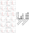
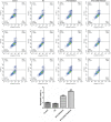
Results: The levels of FITC-labeled IP10-CDR3scfv and IP10-CDR3-PE40scfv proteins in the SKOV3+IP10-CDR3scfv group and the SKOV3+IP10-CDR3-PE40scfv group were remarkably higher than that in the SKOV3 group (P<0.05). So was the HOSEpiC related groups. There was no obvious difference in the levels of IP10, CDR3, Caspase-3, cleaved Caspase-3 and Bcl-2 between the control group and the pcDNA3.1(+) NC group. However, compared with the control group, the levels of Caspase-3 and Bcl-2 were reduced notably and the levels of IP10, CDR3 and cleaved Caspase-3 were elevated sharply in the IP10-CDR3scfv and IP10-CDR3-PE40scfv groups (P<0.05). The control group and the pcDNA3.1(+) NC group demonstrated similar cell viability and apoptosis. However, compared with the control group, cell viability in the IP10-CDR3scfv and IP10-CDR3-PE40scfv groups decreased significantly and cell apoptosis increased (P<0.05).
Conclusion: IP10-CDR3 could reduce the viability and induce the apoptosis of ovarian cancer cells by down-regulating the expression of Bcl-2 and Caspase-3.
Keywords: Bcl-2; CDR3; Caspase-3; IP10; cell apoptosis; ovarian cancer.
© 2019 Chen et al.
Conflict of interest statement
The authors report no conflicts of interest in this work.
Figures


References
-
- Qin M, Jin Y, Ma L, et al. The role of neoadjuvant chemotherapy followed by interval debulking surgery in advanced ovarian cancer: a systematic review and meta-analysis of randomized controlled trials and observational studies. Oncotarget. 2018;9:8614–8628. doi: 10.18632/oncotarget.v9i9 - DOI - PMC - PubMed
LinkOut - more resources
Full Text Sources
Research Materials
Miscellaneous

