. 2020 Jan 14:13:87.
doi: 10.3389/fnsys.2019.00087. eCollection 2019.

Fimbria-Fornix Volume Is Associated With Spatial Memory and Olfactory Identification in Humans

Affiliations

Fimbria-Fornix Volume Is Associated With Spatial Memory and Olfactory Identification in Humans

Louisa Dahmani et al. Front Syst Neurosci. .

Abstract

White matter pathways that surround the hippocampus comprise its afferent and efferent connections, and are therefore crucial in mediating the function of the hippocampus. We recently demonstrated a role for the hippocampus in both spatial memory and olfactory identification in humans. In the current study, we focused our attention on the fimbria-fornix white matter bundle and investigated its relationship with spatial memory and olfactory identification. We administered a virtual navigation task and an olfactory identification task to 55 young healthy adults and measured the volume of the fimbria-fornix. We found that the volume of the right fimbria-fornix and its subdivisions is correlated with both navigational learning and olfactory identification in those who use hippocampus-based spatial memory strategies, and not in those who use caudate nucleus-based navigation strategies. These results are consistent with our recent finding that spatial memory and olfaction rely on similar neural networks and structures.

Keywords: fimbria-fornix; hippocampus; navigation; olfaction; spatial memory; white matter.


Figures

Figure 1
Figure 1
The 4-on-8 Virtual Maze. The 4-on-8 Virtual Maze (4/8 VM) consists in an 8-arm radial maze surrounded by landmarks. In Part 1, four of the paths are blocked and four are open. Participants have to retrieve objects at the end of the open paths. In Part 2, the barriers are removed. Participants have to avoid the paths they visited in Part 1 to retrieve the remaining objects. They can learn the object locations using a spatial memory strategy (e.g., “the path is to the left of the boulder”) or a stimulus-response strategy (“From the starting position, I have to take the path straight ahead and then skip a path on the right”). Once participants learn the task to criterion, they are taken to a probe stage, where a wall is raised around the maze that hides the landmarks. People who used a spatial memory strategy during learning make more errors than people who used stimulus-response strategies, as they can no longer use landmarks to find the target paths. At the end of the task, participants have to draw a map of the maze and are administered a verbal report, which serves to determine the strategy they used as well as the number of landmarks they used (e.g., “I used the rock and the tree to find the objects”) and noticed (e.g., “I saw a mountain but I did not use it”).
Figure 2
Figure 2
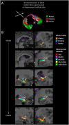
White matter segmentation. (A) 3D reconstruction of the white matter atlas (Amaral et al., 2018), showing the alveus, fimbria, and fornix, superimposed on the hippocampal subfield atlas (Pipitone et al., 2014). (B) Segmentations of the alveus (in orange), fimbria (in blue), and fornix (in purple) are shown for a representative participant. The other colors represent the various hippocampal subfields (not discussed in this article but see Dahmani et al., 2018). Sagittal views are shown on the left, coronal views are shown on the right. A: anterior; P: posterior; R: right; L: left; CA: cornu ammonis; DG: dentate gyrus; SR/SL/SM: stratum radiatum, lacunosum, and moleculare.
Figure 3
Figure 3
Right fimbria-fornix volume is associated with fewer errors during navigational learning in spatial learners, but not response learners. Within spatial learners, average navigational learning errors correlate negatively with right fimbria-fornix volume (shown on the left), r = −0.42 [Bootstrap BCa 95% CI (−0.66, −0.18)], but not with left fimbria-fornix volume (Bootstrap BCa 95% CI cross 0). Within the left fimbria-fornix sub-regions, average navigational learning errors show a marginally significant negative correlation with the left fimbria, r = −0.31 [Bootstrap BCa 95% CI (−0.60, 0.004)], but not with the other sub-regions (Bootstrap BCa 95% CI cross 0). Within response learners, average navigational learning errors do not correlate with left or right fimbria-fornix volume (shown on the right is the right fimbria-fornix volume correlation for comparison, r = 0.10 [Bootstrap BCa 95% CI (−0.26, 0.37)], or any of their sub-regions (Bootstrap BCa 95% CI cross 0). These results indicate that more efficient spatial learning is mainly associated with a greater volume of the right fimbria-fornix fiber system. Stimulus-response learning does not show any correlations with the left or right fimbria-fornix system. †Indicates two overlapping data points.
Figure 4
Figure 4
Olfactory identification correlates positively with right fimbria volume within spatial learners only. Within spatial learners, olfactory identification correlates positively with right fimbria volume (shown on the left), r = 0.41, Bootstrap BCa 95% CI (0.08, 0.68), but not with the left fimbria-fornix or any of its sub-regions (Bootstrap BCa 95% CI cross 0). Within response learners, olfactory identification does not correlate positively with either left or right fimbria-fornix volume or any of their sub-regions {shown on the right is the association with right fimbria volume for comparison, r = 0.18 [Bootstrap BCa 95% CI (−0.07, 0.41)]; all other Bootstrap BCa 95% CI cross 0}. These results indicate that olfactory identification correlates positively with a sub-region of the right fimbria-fornix fiber system in those who use hippocampal-dependent navigation strategies only. †Indicates two overlapping datapoints.

References

    1. Allison S. L., Fagan A. M., Morris J. C., Head D. (2016). Spatial navigation in preclinical Alzheimer’s disease. J. Alzheimers Dis. 52, 77–90. 10.3233/JAD-150855 - DOI - PMC - PubMed
    1. Amaral R. S., Park M. T. M., Devenyi G. A., Lynn V., Pipitone J., Winterburn J., et al. . (2018). Manual segmentation of the fornix, fimbria, and alveus on high-resolution 3T MRI: application via fully-automated mapping of the human memory circuit white and grey matter in healthy and pathological aging. Neuroimage 170, 132–150. 10.1016/j.neuroimage.2016.10.027 - DOI - PubMed
    1. Barrash J. (1998). A historical review of topographical disorientation and its neuroanatomical correlates. J. Clin. Exp. Neuropsychol. 20, 807–827. 10.1076/jcen.20.6.807.1114 - DOI - PubMed
    1. Bohbot V. D., Corkin S. (2007). Posterior parahippocampal place learning in HM. Hippocampus 17, 863–872. 10.1002/hipo.20313 - DOI - PubMed
    1. Bohbot V. D., Iaria G., Petrides M. (2004). Hippocampal function and spatial memory: evidence from functional neuroimaging in healthy participants and performance of patients with medial temporal lobe resections. Neuropsychology 18, 418–425. 10.1037/0894-4105.18.3.418 - DOI - PubMed

LinkOut - more resources