Review
. 2020 Jan 10:10:2909.
doi: 10.3389/fmicb.2019.02909. eCollection 2019.

Microarray Strategies for Exploring Bacterial Surface Glycans and Their Interactions With Glycan-Binding Proteins

Affiliations
Review

Microarray Strategies for Exploring Bacterial Surface Glycans and Their Interactions With Glycan-Binding Proteins

María Asunción Campanero-Rhodes et al. Front Microbiol. .

Abstract

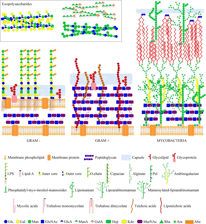
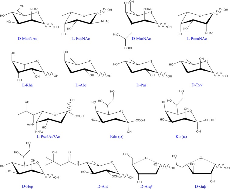
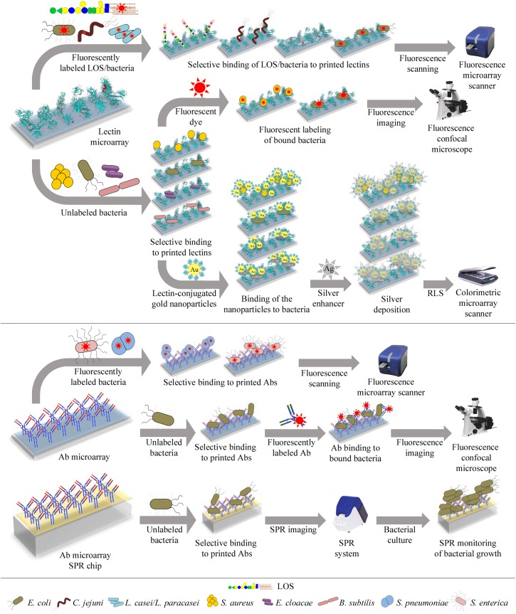
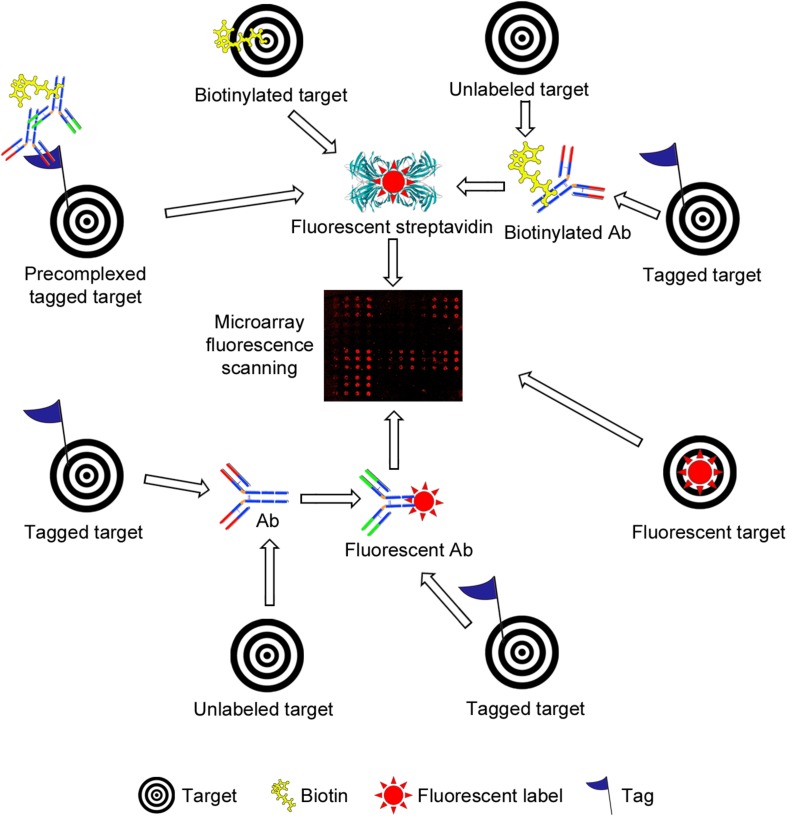
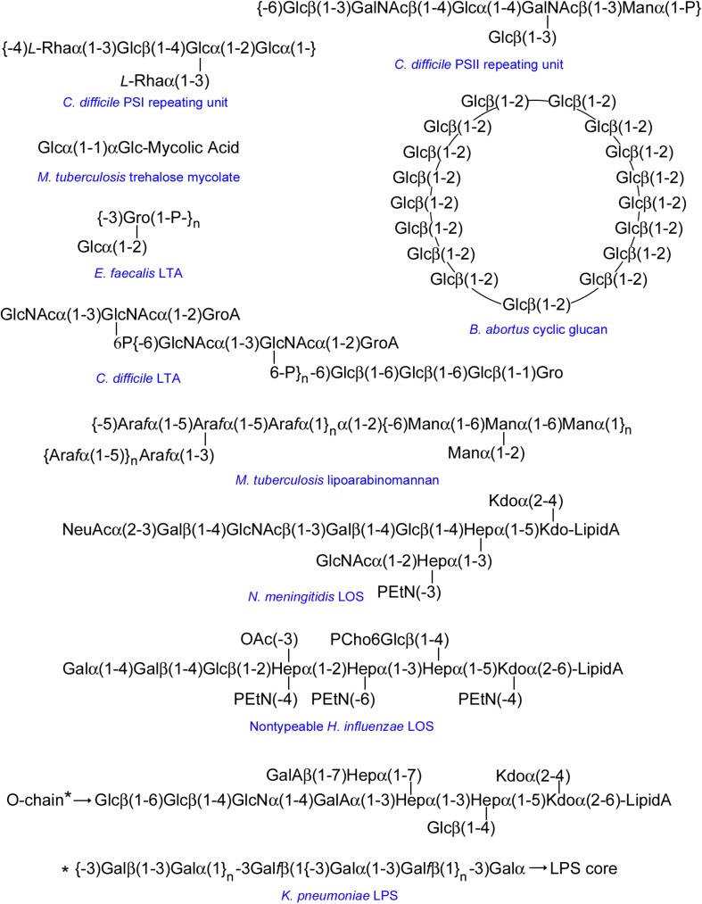
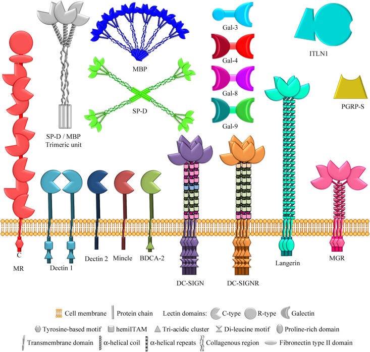
Bacterial surfaces are decorated with distinct carbohydrate structures that may substantially differ among species and strains. These structures can be recognized by a variety of glycan-binding proteins, playing an important role in the bacteria cross-talk with the host and invading bacteriophages, and also in the formation of bacterial microcolonies and biofilms. In recent years, different microarray approaches for exploring bacterial surface glycans and their recognition by proteins have been developed. A main advantage of the microarray format is the inherent miniaturization of the method, which allows sensitive and high-throughput analyses with very small amounts of sample. Antibody and lectin microarrays have been used for examining bacterial glycosignatures, enabling bacteria identification and differentiation among strains. In addition, microarrays incorporating bacterial carbohydrate structures have served to evaluate their recognition by diverse host/phage/bacterial glycan-binding proteins, such as lectins, effectors of the immune system, or bacterial and phagic cell wall lysins, and to identify antigenic determinants for vaccine development. The list of samples printed in the arrays includes polysaccharides, lipopoly/lipooligosaccharides, (lipo)teichoic acids, and peptidoglycans, as well as sequence-defined oligosaccharide fragments. Moreover, microarrays of cell wall fragments and entire bacterial cells have been developed, which also allow to study bacterial glycosylation patterns. In this review, examples of the different microarray platforms and applications are presented with a view to give the current state-of-the-art and future prospects in this field.

Keywords: antibodies; bacterial glycans; bacterial interactions; immune system; lectins; microarrays; vaccine development.


Figures

FIGURE 1
FIGURE 1
Bacterial glycans and architecture of the cell wall of different bacterial groups. Gram-negative bacteria (left part) contain a thin peptidoglycan layer, sandwiched between two cell membranes, and display LPSs (composed of lipid A, inner and outer core, and O-chain) anchored to the outer membrane. Gram-positive bacteria (middle part) contain a thick peptidoglycan layer, covering the cell membrane, and usually display teichoic acids anchored to the membrane (lipoteichoic acids) or bound to the peptidoglycan. Gram-negative and -positive bacteria may also present cell surface glycolipids, glycoproteins, and a polysaccharide capsule. Moreover, they may also secret different polysaccharides (known as exopolysaccharides) into the external environment. Representative exopolysaccharide structures of cepacian (produced by B. cepacia), alginate, and Psl (produced by P. aeruginosa) are shown in the inset. Mycobacteria (right part) contain a large cell wall complex formed by peptidoglycan, arabinogalactan, and mycolic acids of the so-called mycomembrane, and display other distinctive glycan structures, such as lipomannan, lipoarabinomannan, phosphatidyl-myo-inositol-mannosides, and trehalose mycolates. Sugar residues are depicted using the Symbol Nomenclature for Glycans (SNFG) (Varki et al., 2015; Neelamegham et al., 2019).
FIGURE 2
FIGURE 2
Monosaccharide residues found in bacteria, but not in mammals. Only those monosaccharides mentioned in the text have been included. ManNAc, N-acetyl-mannosamine; FucNAc, N-acetyl-fucosamine; MurNAc, N-acetyl-muramic acid; PneuNAc; N-acetyl-pneumosamine; Rha, rhamnose; Abe, abequose; Par, paratose; Tyv, tyvelose; Pse5Ac7Ac, 5,7-di-N-acetyl pseudaminic acid; Kdo, 3-deoxy-D-manno-oct-2-ulosonic acid; Ko, D-glycero-D-talo-oct-2-ulosonic acid; Hep, L-glycero-D-mannoheptose; Ant, anthrose; Araf, arabinofuranose; Galf, galactofuranose.
FIGURE 3
FIGURE 3
Illustration of different lectin and antibody microarray approaches used for glycophenotyping, differentiation, and detection of bacteria. Top panel: Microarrays containing a collection of lectins with diverse carbohydrate-binding specificities can be incubated with fluorescently-labeled bacteria or LOS and bound targets directly quantified using a fluorescence microarray scanner (upper row). Alternatively, the microarrays can be incubated with unlabeled bacteria and bound bacteria next labeled with a fluorescent dye, enabling detection by confocal microscopy (middle row). Bound unlabeled bacteria can also be detected by incubation with gold nanoparticles conjugated to a lectin known to recognize the bacteria under study. The resonance light scattering (RLS) of the nanoparticles is then enhanced by deposition of silver and next measured using a colorimetric microarray scanner (lower row). Bottom panel: Microarrays containing antibodies (Abs) raised against selected bacteria can be incubated with fluorescently-labeled bacteria and bound bacteria detected by fluorescence scanning (upper row). The microarrays can also be incubated with unlabeled bacteria, followed by incubation with fluorescently-labeled anti-bacteria Abs, and bound Abs are next detected by confocal microscopy (middle row). Finally, bacteria selectively bound by Abs arrayed on SPR (surface plasmon resonance) chips can be detected by monitoring their growth during on-chip culture, using SPR imaging (lower row). Specific bacteria that have been tested using these different approaches are detailed in each case.
FIGURE 4
FIGURE 4
Schematic representation of different strategies used for fluorescence-based detection of lectin and antibody (Ab) binding to bacterial carbohydrate and whole cell microarrays. The simplest setup involves incubation with a fluorescently labeled target (lower right side). A common strategy is the use of biotinylated targets, which are next detected by incubation with fluorescently labeled streptavidin (upper part). The targets may carry other tags (e.g., His- or Fc-tags), and detection then has involved the use of biotinylated or unlabeled Abs, followed by incubation with streptavidin or with a biotinylated secondary Ab, as appropriate. Pre-complexing tagged targets with primary and secondary Abs has been exploited to reduce the number of incubation steps and/or to increase the sensitivity of detection. Alternatively, tagged targets have been detected by incubation with a fluorescent or unlabeled Ab, the latter followed by incubation with a fluorescent secondary Ab (lower part). Finally, the binding of unlabeled targets has been monitored using biotinylated or unlabeled primary Abs followed by fluorescently labeled streptavidin or secondary Abs. In all cases, the final step involves the scanning of fluorescence signals.
FIGURE 5
FIGURE 5
Selected examples of representative structures printed in bacterial carbohydrate microarrays. The repeating unit of Enterococcus faecalis LTA is shown as representative of a glycerol phosphate LTA backbone. Bacterial β(1-2)-linked cyclic glucans can occur in unsubstituted form or substituted at Glc C6 with anionic groups (e.g., succinyl in B. abortus). The structure shown for NTHi LOS corresponds to the major glycoform of strain 375. Because of space limitations, the structure of the O-chain of K. pneumoniae LPS is shown below the structure of the LPS core. Unless specifically indicated, sugar units are D-stereoisomers in pyranoside form. Sugars in furanoside form are labeled with the f suffix. GroA, glyceric acid; Gro, glycerol; GalA, galacturonic acid; P, phosphate; PEtN, phosphoethanolamine; OAc, O-acetyl; PCho, phosphorylcholine.
FIGURE 6
FIGURE 6
Lectins of the innate immune system examined using bacterial glycan microarrays. The lectins studied comprise several multimodular membrane receptors that contain C-type carbohydrate-recognition domains (Solís et al., 2015) in addition to other non-lectin domains, specified in each case. The mannose receptor also contains an R-type lectin domain. Soluble lectins examined also include two C-type lectins of the collectin family (so called because they contain a collagenous region), which form different multimeric structures based on similar trimeric units. Other soluble lectins studied are different members of the galectin family, belonging to the chimera type (containing one carbohydrate-recognition domain) and tandem-repeat type (containing two different carbohydrate-recognition domains) structural subgroups, intelectin-1 (a member of the X-type lectin family), and a peptidoglycan recognition protein. MR, mannose receptor; BDCA-2, blood dendritic cell antigen 2; DC-SIGN, dendritic cell-specific ICAM-3-grabbing nonintegrin; DC-SIGNR, endothelial cell DC-SIGN homolog; MGR, macrophage galactose receptor; SP-D, surfactant protein D; MBP, serum mannose-binding protein; Gal-3/4/8/9, galectins 3/4/8/9; ITLN1, intelectin-1; PGRP-S, short peptidoglycan recognition protein.

Similar articles

Cited by

References

    1. Adibekian A., Stallforth P., Hecht M. L., Werz D. B., Gagneux P., Seeberger P. H. (2011). Comparative bioinformatics analysis of the mammalian and bacterial glycomes. Chem. Sci. 2 337–344. 10.1093/glycob/cwu092 - DOI - PubMed
    1. Angata T., Ishii T., Motegi T., Oka R., Taylor R. E., Soto P. C., et al. (2013). Loss of Siglec-14 reduces the risk of chronic obstructive pulmonary disease exacerbation. Cell. Mol. Life Sci. 70 3199–3210. 10.1007/s00018-013-1311-7 - DOI - PMC - PubMed
    1. Angenendt P. (2005). Progress in protein and antibody microarray technology. Drug Discov. Today 10 503–511. 10.1016/S1359-6446(05)03392-1 - DOI - PubMed
    1. Blanchard B., Nurisso A., Hollville E., Tetaud C., Wiels J., Pokorna M., et al. (2008). Structural basis of the preferential binding for globo-series glycosphingolipids displayed by Pseudomonas aeruginosa lectin I. J. Mol. Biol. 383 837–853. 10.1016/j.jmb.2008.08.028 - DOI - PubMed
    1. Blanco Y., Prieto-Ballesteros O., Gómez M. J., Moreno-Paz M., García-Villadangos M., Rodríguez-Manfredi J. A., et al. (2012). Prokaryotic communities and operating metabolisms in the surface and the permafrost of Deception Island (Antarctica). Environ. Microbiol. 14 2495–2510. 10.1111/j.1462-2920.2012.02767.x - DOI - PubMed

LinkOut - more resources