. 2020 Jan 15:10:3054.
doi: 10.3389/fmicb.2019.03054. eCollection 2019.

Molecular Evolution of the Fusion Protein (F) Gene in Human Respirovirus 3

Affiliations

Molecular Evolution of the Fusion Protein (F) Gene in Human Respirovirus 3

Jumpei Aso et al. Front Microbiol. .

Abstract

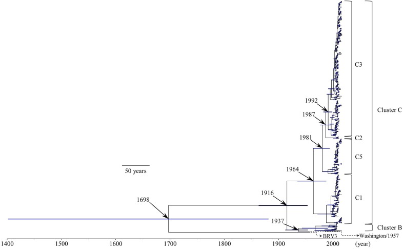
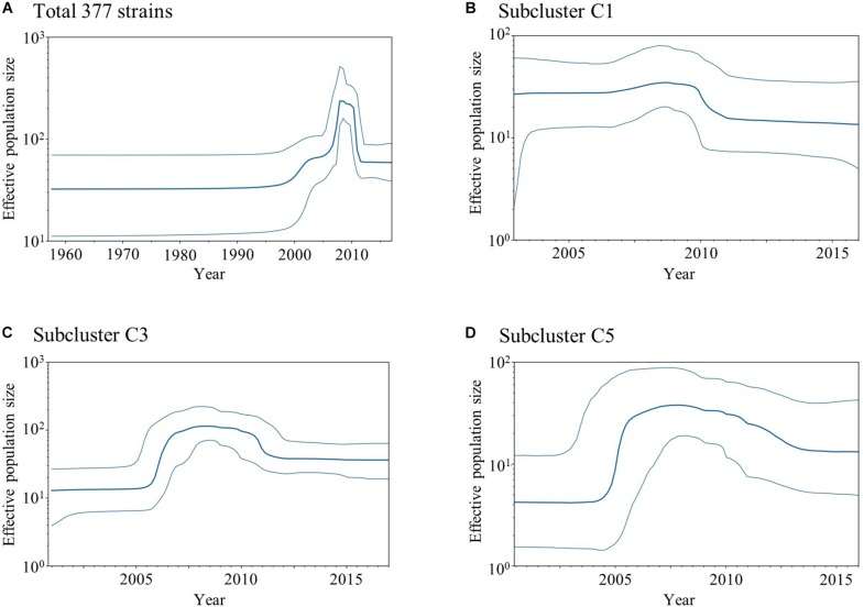
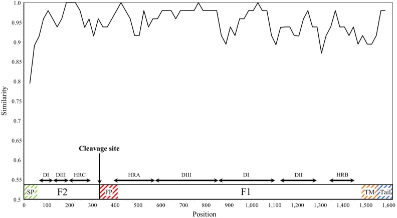
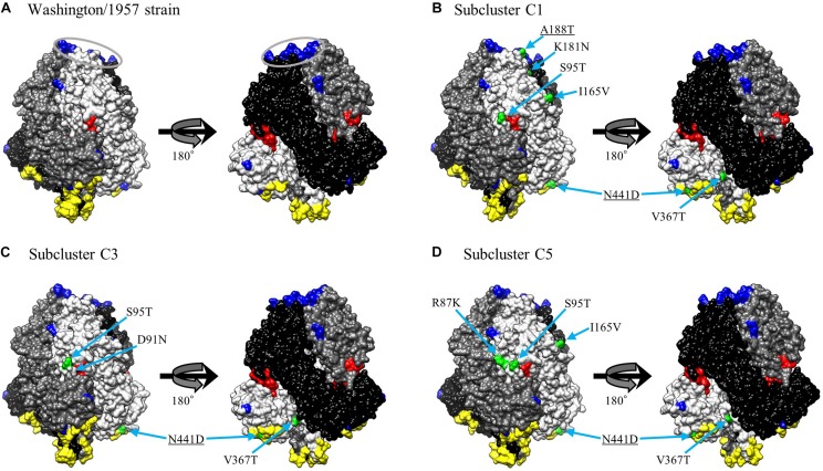
To elucidate the evolution of human respirovirus 3 (HRV3), we performed detailed genetic analyses of the F gene (full-length) detected from hundreds of HRV3 strains obtained from various geographic regions. First, we performed time-scaled evolutionary analyses using the Bayesian Markov chain Monte Carlo method. Then, we performed analyses of phylodynamics, similarity, phylogenetic distance, selective pressure, and conformational B-cell epitope with the F-protein structural analyses. Time-scaled phylogenetic tree showed that the common ancestor of HRV3 and bovine respirovirus 3 diverged over 300 years ago and subdivided it into three major clusters and four subclusters during the most recent 100 years. The overall evolutionary rate was approximately 10-3 substitutions/site/year. Indigenous similarity was seen in the present strains, and the mean phylogenetic distance were 0.033. Many negative selection sites were seen in the ectodomain. The conformational epitopes did not correspond to the neutralizing antibody binding sites. These results suggest that the HRV3 F gene is relatively conserved and restricted in this diversity to preserve the protein function, although these strains form many branches on the phylogenetic tree. Furthermore, HRV3 reinfection may be responsible for discordances between the conformational epitopes and the neutralizing antibody binding sites of the F protein. These findings contribute to a better understanding of HRV3 virology.

Keywords: conformational epitope; fusion peptide; fusion protein (F) gene; human respirovirus 3; molecular evolution.


Figures

FIGURE 1
FIGURE 1
Time-scaled evolutionary tree of the full-length HRV3 F gene constructed by the Bayesian MCMC method. The scale bar represents time (year). Blue bars indicate the 95% highest posterior density (HPD) for each branch year.
FIGURE 2
FIGURE 2
Plots of BSP analyses for the HRV3 F gene. Each panel illustrates the phylodynamics of all 377 strains (A), subcluster C1 (B), subcluster C3 (C), and subcluster C5 (D). Y and x-axes indicate the effective population size and time in years, respectively. The thick blue line shows the median value over time. 95% HPD intervals are represented by thin blue lines.
FIGURE 3
FIGURE 3
Plot of similarity analysis of the F gene across all HRV3 strains. Nucleotide similarity to the prototype strain (Washington/1957 strain) was evaluated using SimPlot. Nucleotide position numbers correspond to the F gene in the prototype strain. The cleavage site and the positions of each domains for the F1 and F2 subunits are shown below the graph (Yin et al., 2005). SP, signal peptide; DI–DIII, domains I–III; HRA–HRC, heptad repeat A–C; FP, fusion peptide; TM, transmembrane anchor; and Tail, cytoplasmic tail.
FIGURE 4
FIGURE 4
Distribution of phylogenetic distances between the full-length sequences of the F gene of all HRV3 strains. The y-axis and x-axis indicate the number of sequence pairs and phylogenetic distances, respectively. Each colored bar represents the pairs to which the strains belonging: gray, different clusters; orange, same cluster; and blue, same subcluster.
FIGURE 5
FIGURE 5
Structural models of the prefusion F protein of Washington/1957 strain (A), subcluster C1 (B), subcluster C3 (C), and subcluster C5 (D). Chains of the trimeric structures are colored in light gray (chain A), dim gray (chain B), and black (chain C). The fusion peptide is shown in red. Conformational epitopes of each strain are indicated in yellow. Previously identified epitopes (MAb binding sites and FAb binding sites) are indicated in blue with the FAb binding sites circled in gray. Amino acid substitution sites of chain A for each variant strain relative to the prototype strain are shown in green. The underlined substitution sites represent the overlap with MAb or FAb binding sites.

References

    1. Abedi G. R., Prill M. M., Langley G. E., Wikswo M. E., Curns A. T., Schneider E. (2016). Estimates of parainfluenza virus-associated hospitalizations and cost among children aged less than 5 years in the United States, 1998-2010. J. Pediatric Infect. Dis. Soc. 5 7–13. 10.1093/jpids/piu047 - DOI - PMC - PubMed
    1. Boskova V., Bonhoeffer S., Stadler T. (2014). Inference of epidemiological dynamics based on simulated phylogenies using birth-death and coalescent models. PLoS Comput. Biol. 10:e1003913. 10.1371/journal.pcbi.1003913 - DOI - PMC - PubMed
    1. Bouckaert R., Heled J., Kühnert D., Vaughan T., Wu C. H., Xie D., et al. (2014). BEAST 2: a software platform for bayesian evolutionary analysis. PLoS Comput. Biol. 10:e1003537. 10.1371/journal.pcbi.1003537 - DOI - PMC - PubMed
    1. Chang A., Dutch R. E. (2012). Paramyxovirus fusion and entry: multiple paths to a common end. Viruses 4 613–636. 10.3390/v4040613 - DOI - PMC - PubMed
    1. Coelingh K. V., Winter C. C. (1990). Naturally occurring human parainfluenza type 3 viruses exhibit divergence in amino acid sequence of their fusion protein neutralization epitopes and cleavage sites. J. Virol. 64 1329–1334. - PMC - PubMed

LinkOut - more resources