. 2020 Jan 14:10:3011.
doi: 10.3389/fimmu.2019.03011. eCollection 2019.

Effects of Vitamin D2 (Ergocalciferol) and D3 (Cholecalciferol) on Atlantic Salmon (Salmo salar) Primary Macrophage Immune Response to Aeromonas salmonicida subsp. salmonicida Infection

Affiliations

Effects of Vitamin D2 (Ergocalciferol) and D3 (Cholecalciferol) on Atlantic Salmon (Salmo salar) Primary Macrophage Immune Response to Aeromonas salmonicida subsp. salmonicida Infection

Manuel Soto-Dávila et al. Front Immunol. .

Abstract

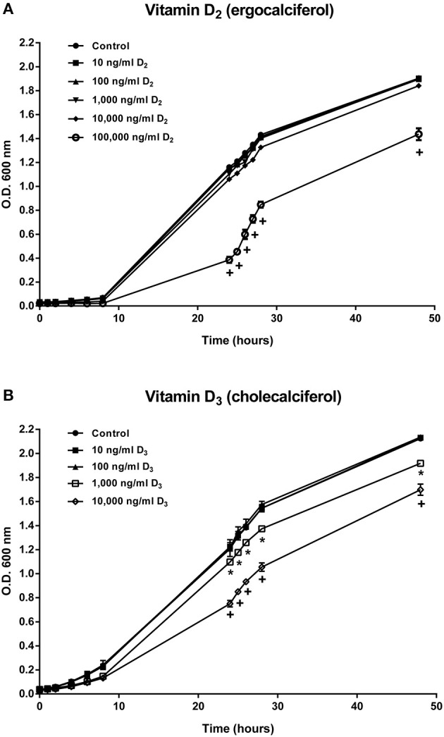
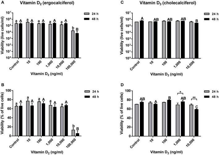
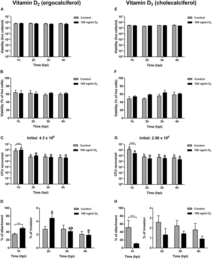
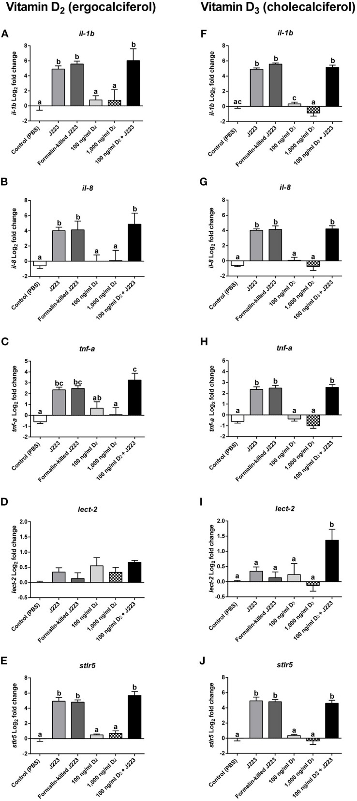
Vitamin D2 (ergocalciferol) and vitamin D3 (cholecalciferol) are fat-soluble secosteroid hormones obtained from plant and animal sources, respectively. Fish incorporates vitamin D2 and D3 through the diet. In mammals, vitamin D forms are involved in mineral metabolism, cell growth, tissue differentiation, and antibacterial immune response. Vitamin D is an essential nutrient in aquafeeds for finfish. However, the influence of vitamin D on fish cell immunity has not yet been explored. Here, we examined the effects of vitamin D2 and vitamin D3 on Salmo salar primary macrophage immune response to A. salmonicida subspecies salmonicida infection under in vitro conditions. We determined that high concentrations of vitamin D2 (100,000 ng/ml) and D3 (10,000 ng/ml) affect the growth of A. salmonicida and decrease the viability of S. salar primary macrophages. In addition, we determined that primary macrophages pre-treated with a biologically relevant concentration of vitamin D3 for 24 h showed a decrease of A. salmonicida infection. In contrast, vitamin D2 did not influence the antibacterial activity of the S. salar macrophages infected with A. salmonicida. Vitamin D2 and D3 did not influence the expression of canonical genes related to innate immune response. On the other hand, we found that A. salmonicida up-regulated the expression of several canonical genes and suppressed the expression of leukocyte-derived chemotaxin 2 (lect-2) gene, involved in neutrophil recruitment. Primary macrophages pre-treated for 24 h with vitamin D3 counteracted this immune suppression and up-regulated the transcription of lect-2. Our results suggest that vitamin D3 affects A. salmonicida attachment to the S. salar primary macrophages, and as a consequence, the A. salmonicida invasion decreased. Moreover, our study shows that the positive effects of vitamin D3 on fish cell immunity seem to be related to the lect-2 innate immunity mechanisms. We did not identify positive effects of vitamin D2 on fish cell immunity. In conclusion, we determined that the inactive form of vitamin D3, cholecalciferol, induced anti-bacterial innate immunity pathways in Atlantic salmon primary macrophages, suggesting that its utilization as a component of a healthy aquafeed diet in Atlantic salmon could enhance the immune response against A. salmonicida.

Keywords: Aeromonas salmonicida; Atlantic salmon; Gram-negative; innate immunity; primary macrophages; vitamin D2; vitamin D3.


Figures

Figure 1
Figure 1
Aeromonas salmonicida subsp. salmonicida growth curve in TSB media supplemented with (A) 10, 100, 1,000, 10,000, and 100,000 ng ml−1 of vitamin D2 and (B) 10, 100, 1,000, and 10,000 ng ml−1of vitamin D3. Growth was determined by reading O.D. 600 nm at different time points until 48 h. Each value is the mean ± S.E.M (n = 3). Symbols (*, +) indicate differences between each group at different time points of measure, p < 0.05.
Figure 2
Figure 2
Atlantic salmon primary macrophages treated with vitamin D2 or D3. (A) Live cells and (B) percentage of viability of primary macrophages treated with 10, 100, 1,000, 10,000, and 100,000 of vitamin D2, were measured after 24 and 48 h of treatment. (C) Live cells and (D) percentage of viability of primary macrophages treated with 10, 100, 1,000, and 10,000 ng ml−1 of vitamin D3 were measured after 24 and 48 h of treatment. Each value represents the mean ± S.E.M (n = 6). Lower case letters (a, b) show differences between treatments after 24 h. Upper case letters (A,B,C) show differences between treatments after 48 h. Asterisks (*) represent significant differences between treatments (*p < 0.05, **p < 0.01).
Figure 3
Figure 3
Gentamicin exclusion assay in Atlantic salmon primary macrophages pre-treated for 24 h with either control or 100 ng ml−1 of vitamin D2 or vitamin D3, and then infected with Aeromonas salmonicida subsp. salmonicida. Live cells of primary macrophages pre-treated with vitamin D2 (A), or vitamin D3 (E); percentage of viability of primary macrophages pre-treated with vitamin D2 (B), or vitamin D3 (F), Colony forming unit (CFU) of A. salmonicida in Atlantic salmon primary macrophages pre-treated with vitamin D2 (C), or vitamin D3 (G); and percentage of attachment and invasion of A. salmonicida in Atlantic salmon primary macrophages pre-treated with vitamin D2 (D), or vitamin D3 (H), were measured 1, 2, 3, and 4 h post-infection. Initial A. salmonicida inoculum calculated in TSA Congo red plates are shown above the CFU figures. Each value represents the mean ± S.E.M (n = 6). Asterisks (*) represent significant differences between treatments on each time-point (**p < 0.01, ***p < 0.001). Lower case letters (a, b) show differences in the control at different time points post-infection. Upper case letters (A, B) show differences in vitamin D2 or D3 pre-treated cells in different time points post-infection.
Figure 4
Figure 4
Gene expression of (A,F) Interleukin 1b (il-1b), (B,G) Interleukin 8 (il-8), (C,H) Tumor necrosis factor alpha (tnf-α), (D,I) Leukocyte-derived chemotaxin 2 (lect-2), and (E,J) soluble toll-like receptor 5 (stlr5) in Atlantic salmon primary macrophages isolated from head kidney pre-treated 24 h with either the control, vitamin D2 (100 and 1,000 ng ml−1) or vitamin D3 (100 and 1,000 ng ml−1), and then inoculated with PBS (control) or infected with live (J223) or exposed to formalin-killed A. salmonicida for 3 h. Relative expression was calculated using the 2(−ΔΔCt) method and Log2 converted using β-actin and 60S ribosomal protein L32 (rpl32) as internal reference genes. Each value is the mean ± S.E.M (n = 6). Different letters represent significant differences between treatments, p < 0.05.

Similar articles

Cited by

References

    1. Zittermann A. Vitamin D in preventive medicine: are we ignoring the evidence? Br J Nutr. (2003) 89:552–72. 10.1079/BJN2003837 - DOI - PubMed
    1. Grant W. Epidemiology of disease risks in relation to vitamin D insufficiency. Prog Biophys Mol Bio. (2006) 92:65–79. 10.1016/j.pbiomolbio.2006.02.013 - DOI - PubMed
    1. Lips P. Vitamin D physiology. Prog Biophys Mol Bio. (2006) 92:4–8. 10.1016/j.pbiomolbio.2006.02.016 - DOI - PubMed
    1. Lock EJ, Waagbø R, Wendelaar-Bonga S, Flick G. The significance of vitamin D for fish: a review. Aquac Nutr. (2010) 16:100–16. 10.1111/j.1365-2095.2009.00722.x - DOI
    1. Borges M, Martini L, Rogero M. Current perspectives on vitamin D, immune system, and chronic diseases. Nutrition. (2011) 27:399–404. 10.1016/j.nut.2010.07.022 - DOI - PubMed

Publication types

MeSH terms