. 2020 Jan 17:10:3105.
doi: 10.3389/fimmu.2019.03105. eCollection 2019.

Toxoplasma gondii Recruits Factor H and C4b-Binding Protein to Mediate Resistance to Serum Killing and Promote Parasite Persistence in vivo

Affiliations

Toxoplasma gondii Recruits Factor H and C4b-Binding Protein to Mediate Resistance to Serum Killing and Promote Parasite Persistence in vivo

Patricia M Sikorski et al. Front Immunol. .

Abstract

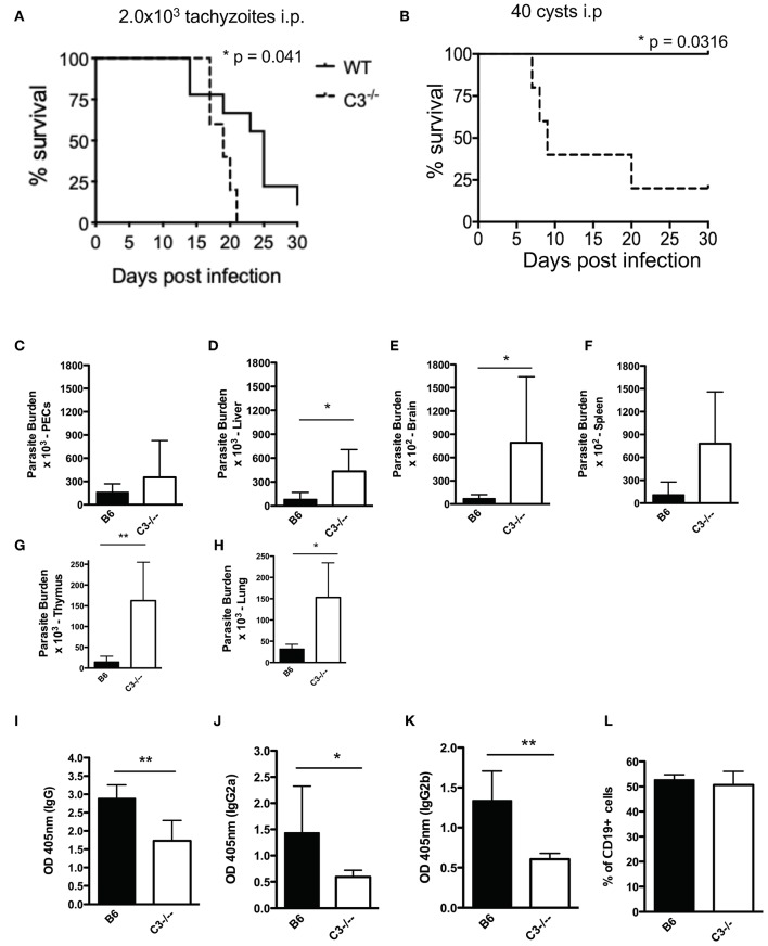
Regulating complement is an important step in the establishment of infection by microbial pathogens. Toxoplasma gondii actively resists complement-mediated killing in non-immune human serum (NHS) by inactivating C3b, however the precise molecular basis is unknown. Here, a flow cytometry-based C3b binding assay demonstrated that Type II strains had significantly higher levels of surface-bound C3b than Type I strains. However, both strains efficiently inactivated C3b and were equally resistant to serum killing, suggesting that resistance is not strain-dependent. Toxoplasma activated both the lectin (LP) and alternative (AP) pathways, and the deposition of C3b was both strain and lectin-dependent. A flow cytometry-based lectin binding assay identified strain-specific differences in the level and heterogeneity of surface glycans detected. Specifically, increased lectin-binding by Type II strains correlated with higher levels of the LP recognition receptor mannose binding lectin (MBL). Western blot analyses demonstrated that Toxoplasma recruits both classical pathway (CP) and LP regulator C4b-binding proteins (C4BP) and AP regulator Factor H (FH) to the parasite surface to inactivate bound C3b-iC3b and C3dg and limit formation of the C5b-9 attack complex. Blocking FH and C4BP contributed to increased C5b-9 formation in vitro. However, parasite susceptibility in vitro was only impacted when FH was blocked, indicating that down regulation of the alternative pathway by FH may be more critical for parasite resistance. Infection of C3 deficient mice led to uncontrolled parasite growth, acute mortality, and reduced antibody production, indicating that both the presence of C3, and the ability of the parasite to inactivate C3, was protective. Taken together, our results establish that Toxoplasma regulation of the complement system renders mice resistant to acute infection by limiting parasite proliferation in vivo, but susceptible to chronic infection, with all mice developing transmissible cysts to maintain its life cycle.

Keywords: Toxoplasma gondii; alternative pathway regulation; complement system resistance; immune evasion; protozoan parasites.


Figures

Figure 1
Figure 1
Strain-specific difference in C3b deposition. (A) Titration of NHS. Type II ME49 parasite were incubated in non-immune human serum (NHS) serially diluted 2-fold, from 40 to 2.5%. Dilutions were made in Hanks Buffered Saline Solution supplemented with 0.15 mM CaCl2 and 1 mM MgCl2 to facilitate classical/lectin and alternative pathway activation, respectively (HBSS++). Parasites were stained with a mouse monoclonal α-human C3b/iC3b antibody, 1:500 (Cedarlane). 10 mM EDTA was used as a negative control to inhibit all complement pathways. C3b deposition was measured by flow cytometry. Flow cytometry data are shown as mean ±SEM from three independently performed experiments. (B) 1 × 10⁶ Type II ME49 (closed circle) and 1 × 10⁶ Type I RH (closed square) parasites were incubated in 10% NHS in HBSS++ over 60 min and C3b deposition was measured using flow cytometry as described above. ME49 parasites incubated with 10% heat inactivated serum (hiNHS) was used as a negative control (open square, dotted line). Flow cytometry data are shown as mean ±SEM from five independently performed experiments and statistically significant differences between Type I and Type II strains were determined using multiple Student's t test with Holm-Sidak correction for multiple comparisons, **p < 0.005, ***p < 0.001. (C) Flow cytometry results from C3b deposition time course comparing Type II strains ME49 (black) and CZ1 (gray) in 10% NHS over 60 min. Flow cytometry data are shown as mean ±SEM from three independently performed experiments; no significant differences between Type II strains CZ1 and ME49 using multiple Student's t test with Holm-Sidak correction for multiple comparisons, ns = not significant. (D) Western blots showing the difference in levels of C3b between 1 × 10⁶ Type II ME49 vs. 1 × 10⁶ RH parasites incubated 20' in 10% NHS at 37°C (E) iC3b/C3b ratio for ME49 and RH incubated in 10% NHS for 20 min. The ratio was determined by quantifying the density of active C3b (using the 75 kDa beta chain of C3b) and inactive iC3b and C3dg (43 and 41 kDa bands, respectively) from three independent western blot images using Image J software. There was no significant difference between the ratio of iC3b/C3b between ME49 and RH using an unpaired Student's t test, ns, not significant.
Figure 2
Figure 2
Serum resistance is strain independent. (A) Kinetics of C3b inactivation on Type II ME49 parasites (5 × 10⁶) over 60 min in 10% NHS probed for C3 and its catabolites using a goat anti-human C3 polyclonal antibody 1:20,000 (CompTech) and donkey anti-goat 1:5,000 (Santa Cruz Biotechnology). Arrows correspond to sizes of C3 (active C3b α chain 110 kDa, β chain 75 kDa; inactive iC3b α1 68 & α2 43 kDa, and C3dg 41 kDa). SRS29B (formerly SAG1, 1:5,000) was used as loading control. Western blot image shown is representative of at least three independent experiments with similar results. Parasite viability and C5b-9 pore formation was determined by exposing parasites to 10, 20, 40, 60% non-immune (black bars) or immune (open bars) serum for 60 min at 37°C. 20% heat inactivated (hiNHS) non-immune serum was used a negative control. Flow cytometry was used to measure parasite susceptibility by staining Type II (B) and Type I (C) parasites with a fixable viability stain (Life Technologies) and membrane attack complex formation (D,E) using monoclonal mouse α-human C5b-9 (Santa Cruz Biotechnology, 1:500). Immune serum was used as a positive control for parasite lysis due to classical pathway activation. Flow cytometry data are shown as mean ± SEM from three independently performed experiments. Statistically significant differences between parasites incubated in immune vs. non-immune serum were determined using multiple Student's t test with Holm-Sidak correction for multiple comparisons, *p < 0.05, **p < 0.01, ***p < 0.001.
Figure 3
Figure 3
Toxoplasma gondii activates Ca2+ dependent complement pathways. Flow cytometry results of C3b deposition time course over 60 min at 37°C on Type II ME49 parasites (A) and Type I RH parasites (B) incubated in 10% NHS in HBSS++ or with HBSS++ treated with 10 mM MgEGTA to inactivate CP and LP. Ten millimolar EDTA was added to 10% NHS to inactivate all complement pathways (open square, dotted line). Flow cytometry data are shown as mean ± SEM from three independently performed experiments. Significant differences in C3b deposition between parasites exposed to NHS and NHS treated with 10 mM MgEGTA was compared using multiple Student's t test with Holm-Sidak correction for multiple comparisons, **p < 0.01, ****p < 0.0001.
Figure 4
Figure 4
Heterogeneity in surface carbohydrate composition between Type I and Type II strains affects MBL recognition. Carbohydrate ligands on the parasite surface were surveyed using a panel of biotinylated lectins with known specificities (Vector Laboratories, α-linked mannose/α-linked glucose (ConA Concanavalin A), N-Acetylglucosamine, GlcNac (WGA, wheat germ agglutinin), galactose (RCA, Ricinus communis agglutinin I) and, N-Acetylgalactosamine, GalNAc (DBA, Dolichos biflorus agglutinin). 1 × 10⁶ parasites were incubated with 5 μg/ml of biotinylated lectin for 1 h on ice, followed by streptavidin-APC (1:1,000) for 30 min, and analyzed by flow cytometry. Representative histograms of lectin binding between Type II (ME49, blue) (A) and Type I (RH, red) (B) compared to unstained control (black, dotted line). (C) Flow cytometry results of MBL (mannose binding lectin) binding to Type II (ME49, black bar) and Type I (RH, open bar) parasites incubated in 10% NHS for 60' on ice. MBL was detected using goat α- human MBL (R&D Systems 1:1,000) followed by anti-goat-APC (1:1,000). Flow cytometry data are shown as mean ±SEM from three independently performed experiments. Significant differences in MBL binding between Type I RH and Type II ME49 was determined using an unpaired t test, *p < 0.05. (D) Type II ME49 parasites were incubated in 10% serum from a patient with a mutation in the human MBL gene (open circle, dotted line), 10% NHS from a MBL sufficient single donor in HBSS++ (solid circle, solid line) or 10% heat inactivated NHS (hiNHS) in HBSS++ (open square, solid line). Flow cytometry data are shown as mean ± SEM from three independently performed experiments. Significant differences in C3b deposition between parasites exposed to MBL sufficient NHS and MBL deficient NHS was compared using multiple Student's t test with Holm-Sidak correction for multiple comparisons, **p < 0.01, ***p < 0.001.
Figure 5
Figure 5
Toxoplasma gondii recruits AP regulator Factor H and CP/LP regulator C4b-binding protein to the parasite surface. 1 × 10⁶ Type II ME49 parasites were incubated in 10% NHS for 0–60 min at 37°C. Western blots (left panels) of C4BP (A) (rabbit α-human C4BP, AssayPro 1:500) and FH (B) (goat α-human Factor H, CompTech 1:20,000) binding. Serum or purified protein was used as a positive control and heat inactivated serum (hiNHS) was used as a negative control. Blots were stripped and re-probed with anti-SRS29B (SAG1) for loading control. Images are from one representative of three independent experiments with similar results. Right panels (A,B) represent flow cytometric assays of C4BP (A) and FH (B) binding to the parasite surface for 0–60 min. Heat inactivated NHS (hiNHS) serum was used as a negative control. Flow cytometry data are shown as mean ± SEM from three independently performed experiments.
Figure 6
Figure 6
Factor H and C4b-binding protein contribute to serum resistance. Factor H (FH) and C4b-binding protein (C4BP) were blocked by pre-incubating 10% NHS with 1:100 or 1:400 dilution of goat α-human FH (CompTech) or rabbit α-human C4BP (AssayPro) for 1 h on ice before adding to 1 × 10⁶ parasites and incubating for 60 min at 37°C. Flow cytometric analysis of C5b-9 formation (A) and parasite viability (B) after 60' in 10% NHS blocked with 1:100 or 1:400 of α-C4BP (gray bars) α-FH (open bars) antibodies. 10% heat inactivated serum (hiNHS) was used a negative control. Flow cytometry data are shown as mean ± SEM from three independently performed experiments. Significant differences between the compared groups was determined using multiple Student's t test with Holm-Sidak correction for multiple comparisons, *p < 0.05, ***p < 0.001, ****p < 0.0001.
Figure 7
Figure 7
C3 contributes to protection against acute T. gondii infection. Survival of 6–8 week old C57BL6 (n = 10) and C3−/− (n = 6) female mice infected with (A) 2.0 × 10³ CZ1 tachyzoites intraperitoneally or (B) 40 ME49 cysts intraperitoneally. Survival rates were compared by log-rank survival analysis of Kaplan-Meier curves, p = 0.041 and p = 0.0316. Mice infected with 40 cysts intraperitoneally were sacrificed 7 days post infection (n = 5). Parasite burdens were determined by counting plaque forming units (PFUs) by plating (C) peritoneal fluid, and homogenates of (D) liver, (E) brain, (F) spleen, (G) thymus, and (H) lungs onto HFF monolayers in 12 well-plates. Total levels of IgG (I), IgG2a (J), and IgG2b (K) 7 days post infection with 40 cysts i.p. were measured by ELISA. (L) Percentage of CD19+ splenocytes in wild type (B6) and C3−/− mice. Data shown as mean ± SEM from one representative of two independently performed experiments. Significant differences between the compared groups was determined using unpaired Student's t test, *p < 0.05, **p < 0.01.

Similar articles

Cited by

References

    1. Noris M, Remuzzi G. Overview of complement activation and regulation. Semin Nephrol. (2013) 33:479–92. 10.1016/j.semnephrol.2013.08.001 - DOI - PMC - PubMed
    1. Morgan BP. The human complement system in health and disease. Ann Rheum Dis. (1998) 57:581–1. 10.1136/ard.57.10.581 - DOI
    1. Evans-Osses I, de Messias-Reason I, Ramirez MI. The Emerging Role of Complement Lectin Pathway in Trypanosomatids: Molecular Bases in Activation, Genetic Deficiencies, Susceptibility to Infection, and Complement System-Based Therapeutics. In: The Scientific World Journal. (2013). Available online at: https://www.hindawi.com/journals/tswj/2013/675898/ (accessed xMay 21, 2019). 10.1155/2013/675898 - DOI - PMC - PubMed
    1. Iida K, Whitlow MB, Nussenzweig V. Amastigotes of Trypanosoma cruzi escape destruction by the terminal complement components. J Exp Med. (1989) 169:881–91. 10.1084/jem.169.3.881 - DOI - PMC - PubMed
    1. Molina-Cruz A, DeJong RJ, Ortega C, Haile A, Abban E, Rodrigues J, et al. . Some strains of Plasmodium falciparum, a human malaria parasite, evade the complement-like system of Anopheles gambiae mosquitoes. PNAS. (2012) 109:E1957–62. 10.1073/pnas.1121183109 - DOI - PMC - PubMed

Publication types

MeSH terms

Substances