. 2020 Jan 15:10:3115.
doi: 10.3389/fimmu.2019.03115. eCollection 2019.

Endogenous T Cell Receptor Rearrangement Represses Aggressive Central Nervous System Autoimmunity in a TcR-Transgenic Model on the Non-Obese Diabetic Background

Affiliations

Endogenous T Cell Receptor Rearrangement Represses Aggressive Central Nervous System Autoimmunity in a TcR-Transgenic Model on the Non-Obese Diabetic Background

Asmita Pradeep Yeola et al. Front Immunol. .

Abstract

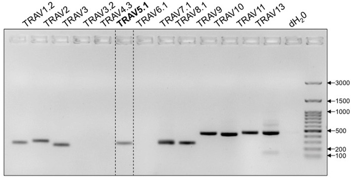
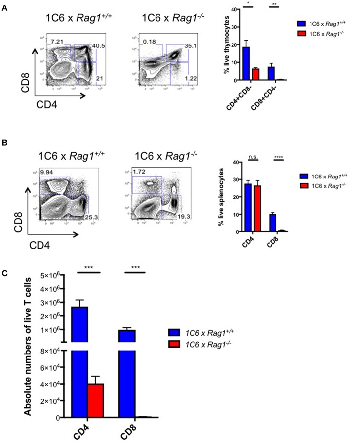
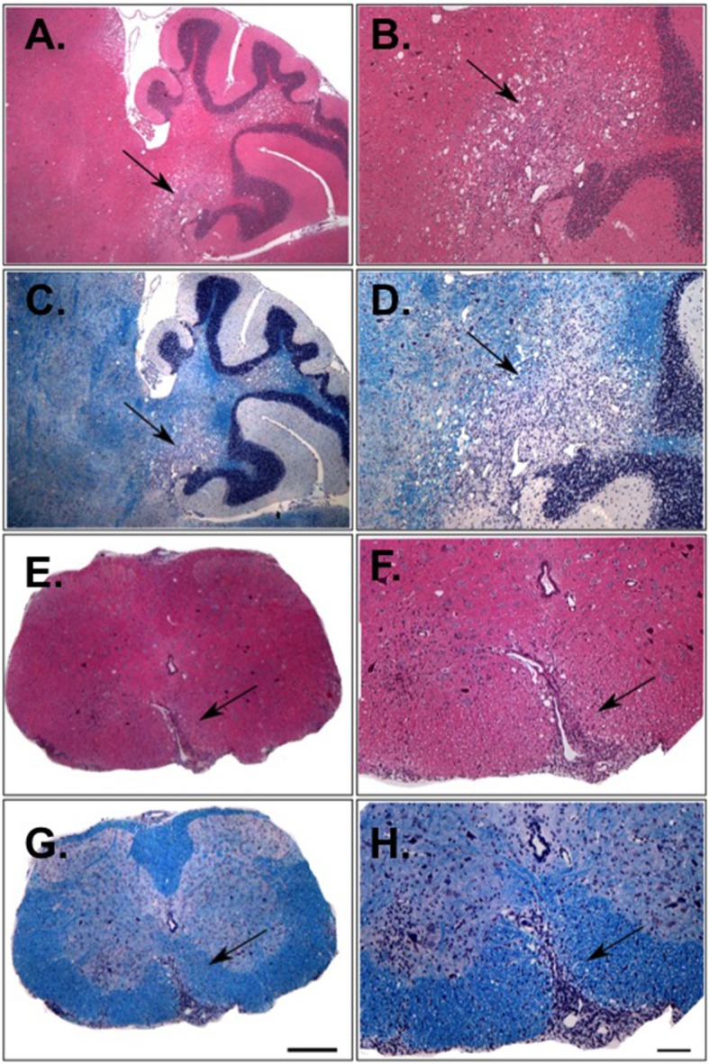
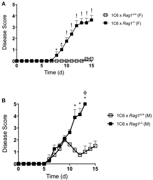
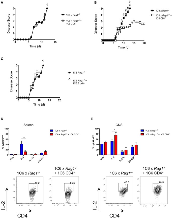
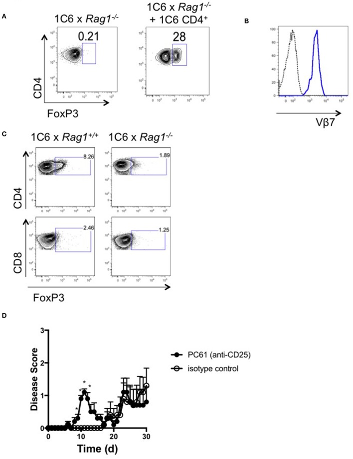
The T cell response to central nervous system (CNS) antigen in experimental autoimmune encephalomyelitis (EAE) permits one to model the immune aspects of multiple sclerosis. 1C6 transgenic mice on the non-obese diabetic (NOD) background possess a class II-restricted T cell receptor (TcR; Vα5-Vβ7) specific for the encephalitogenic peptide myelin oligodendrocyte glycoprotein (MOG)[35-55]. It remains to be determined what role is played by allelic inclusion in shaping the TcR repertoire of these mice. Here, we show that 1C6 T cells display substantial promiscuity in their expression of non-transgenically derived Vα chains. Further, enforced expression of the transgenic TcR in 1C6 × Rag1-/- mice profoundly disrupted thymic negative selection and led to a sharp decrease in the number of mature peripheral T cells. 1C6 × Rag1-/- mice developed spontaneous EAE at a significant frequency and rapidly developed fatal EAE upon immunization with myelin oligodendrocyte glycoprotein (MOG)[35-55]. Passive transfer of 1C6 × Rag1+/+ CD4+ T cells, but not CD8+ T cells or B cells, partially rescued 1C6 × Rag1-/- mice from severe EAE. FoxP3+ CD4+ Treg cells were present in the CNS of immunized 1C6 mice, as well as immunized 1C6 × Rag1-/- that had been supplemented with 1C6 CD4+ T cells. However, they were not observed in 1C6 × Rag1-/- that did not receive Rag1-sufficient 1C6 CD4+. Further, in vivo blockade of Treg accelerated the onset of symptoms in 1C6 mice immunized with MOG[35-55], indicating the pertinence of Treg-mediated control of autoimmune inflammation in this model. Thus, TcR allelic inclusion is crucial to the generation of FoxP3+ CD4+ T cells necessary for the suppression of severe CNS autoimmunity.

Keywords: 1C6; EAE (experimental autoimmune encephalomyelitis); FoxP3; RAG; TCR transgenic mice; Treg—regulatory T cell; allelic exclusion; non-obese diabetic (NOD).


Figures

Figure 1
Figure 1
1C6 Tg T cells are promiscuous in their expression of endogenous TcRα. Purified splenic 1C6 CD8+ T cells were assessed for the presence of specific TcRα DNA by PCR. Transgenically encoded Vα5.1 bracketed by dotted lines. Representative of three experiments.
Figure 2
Figure 2
Peripheral T cell populations are dysregulated in 1C6 × Rag1−/− mice due to defective negative selection. Thymic (A) and splenic (B,C) cell populations were enumerated from female 1C6 (n = 6) vs. 1C6 × Rag1−/− (n = 6) mice. (A) The proportion of thymocytes expressing CD4 and/or CD8 was enumerated. The proportion (B) and absolute frequency (C) of splenic CD4+ and CD8+ T cells were assessed by flow cytometry. Graphs: *p < 0.05; **p < 0.01; ***p < 0.001; ****p < 0.0001, two-tailed t-test. n.s., not significant.
Figure 3
Figure 3
Histological analysis of the CNS of 1C6 × Rag1−/− mice that develop spontaneous EAE. Cerebellar (A–D) and spinal cord (E–H) lesions from an 18-weeks old male 1C6 × Rag1−/− mouse that spontaneously developed paralytic disease. H&E staining (A,B,E,F) was used to identify inflammatory foci, and Luxol fast blue was used to detect myelin (C,D,G,H). Arrows indicate inflammatory damage. (A,C,E,G), 10× magnification. (B,D,F,H), 4× magnification. Scale bars, 100 μm. Representative of eight animals (6 female, 2 male).
Figure 4
Figure 4
1C6 × Rag1−/− mice develop severe MOG[35−55]-driven EAE. 1C6 (male, n = 4; female, n = 5) and 1C6 × Rag1−/− (male, n = 4; female, n = 5) mice were actively immunized with MOG[35−55] and were monitored for signs of EAE. (A) female, (B) male. *p < 0.05; !p < 0.001, two-tailed Mann-Whitney U test. ϕ indicates that all mice in the experimental group attained ethical endpoints. Representative of three immunizations.
Figure 5
Figure 5
1C6 CD4+ T cells restrain CNS autoimmunity in 1C6 × Rag1−/− mice. (A) Male 1C6 × Rag1−/− mice were reconstituted (n = 5), or not (n = 5), with 2 × 10⁶ CD4+ T cells from unmanipulated male 1C6 mice. After 7 days, mice were actively immunized with MOG[35−55] and were monitored for signs of EAE. Representative of 2 experiments. (B) Male 1C6 × Rag1−/− mice were reconstituted (n = 5), or not (n = 5), with 2 × 10⁶ CD8+ T cells from unmanipulated male 1C6 mice. After 7 days, mice were actively immunized with MOG[35−55] and were monitored daily for signs of EAE. *p < 0.05, two-tailed Mann-Whitney U test. ϕ indicates that all mice in the experimental group attained ethical endpoints. (C) Female 1C6 × Rag1−/− mice were reconstituted (n = 4), or not (n = 4), with 2 × 10⁶ CD19+ B cells from unmanipulated 1C6 mice. After 7 days, mice were actively immunized with MOG[35−55] and were monitored for signs of EAE. ϕ indicates that all mice in the experimental group attained ethical endpoints. *p < 0.05; #p < 0.01, two-tailed Mann-Whitney U test. ϕ indicates that all mice in the experimental group attained ethical endpoints. (D,E). Spleen (D) or CNS-infiltrating (E) CD4+ T cells were isolated from immunized 1C6 × Rag1−/− mice when they reached ethical endpoints (n = 9), or from 1C6 × Rag1−/− (reconstituted with 1C6 CD4+) that were sacrificed in parallel (n = 4). T cells were assessed ex vivo for the indicated cytokines by flow cytometry. All data are gated on live CD4+ events. *p < 0.05; **p < 0.01, Sidak's multiple comparisons test after two-way ANOVA. Bottom plots, representative IL-2 expression from splenic (D) or CNS-infiltrating (E) CD4+ T cells.
Figure 6
Figure 6
FoxP3+ CD4+ T cells are absent from the CNS of 1C6 × Rag1−/− mice with EAE. (A,B). Female 1C6 × Rag1−/− mice were reconstituted, or not, with 2 × 10⁶ CD4+ T cells from unmanipulated 1C6 mice. After 14 days, mice were actively immunized with MOG[35−55]. At experimental endpoints, CNS-infiltrating CD4+ T cells were assessed by flow cytometry for expression of FoxP3 (A). Representative data, n = 4 each group. (B) Vβ7 expression (blue line) on CNS-infiltrating CD4+FoxP3+ T cells from a 1C6 × Rag1−/− mouse reconstituted with 1C6 CD4+ T cells. Dotted line, FMO control. (C) Female 1C6 and 1C6 × Rag1−/− mice were actively immunized with MOG[35−55]. Lymph node T cells were isolated at the peak of disease and assessed by flow cytometry for FoxP3 expression. Gated on CD4+ (top) or CD8+ (bottom) events. Representative of 5 mice each. (D) Male 1C6 mice were treated with Treg-depleting anti-CD25 antibody (PC61) or with isotype control on days−11,−9, and−7, and were immunized with MOG[35−55] on d0. n = 5, both groups. *p < 0.05 on individual days, Mann-Whitney U test.

Similar articles

Cited by

References

    1. Haghikia A, Hohlfeld R, Gold R, Fugger L. Therapies for multiple sclerosis: translational achievements and outstanding needs. Trends Mol Med. (2013) 19:309–19. 10.1016/j.molmed.2013.03.004 - DOI - PubMed
    1. Vargas DL, Tyor WR. Update on disease-modifying therapies for multiple sclerosis. J Investig Med. (2017) 65:883–91. 10.1136/jim-2016-000339 - DOI - PubMed
    1. Rangachari M, Kuchroo VK. Using EAE to better understand principles of immune function and autoimmune pathology. J Autoimmun. (2013) 45:31–9. 10.1016/j.jaut.2013.06.008 - DOI - PMC - PubMed
    1. Bettelli E. Building different mouse models for human MS. Ann N Y Acad Sci. (2007) 1103:11–18. 10.1196/annals.1394.021 - DOI - PubMed
    1. Basso AS, Frenkel D, Quintana FJ, Costa-Pinto FA, Petrovic-Stojkovic S, Puckett L, et al. . Reversal of axonal loss and disability in a mouse model of progressive multiple sclerosis. J Clin Invest. (2008) 118:1532–43. 10.1172/JCI33464 - DOI - PMC - PubMed

Publication types

MeSH terms

Substances