. 2020 Feb;19(2):972-980.
doi: 10.3892/etm.2019.8280. Epub 2019 Dec 4.

Enalapril attenuates endoplasmic reticulum stress and mitochondrial injury induced by myocardial infarction via activation of the TAK1/NFAT pathway in mice

Affiliations

Enalapril attenuates endoplasmic reticulum stress and mitochondrial injury induced by myocardial infarction via activation of the TAK1/NFAT pathway in mice

Xing Rong et al. Exp Ther Med. 2020 Feb.

Abstract

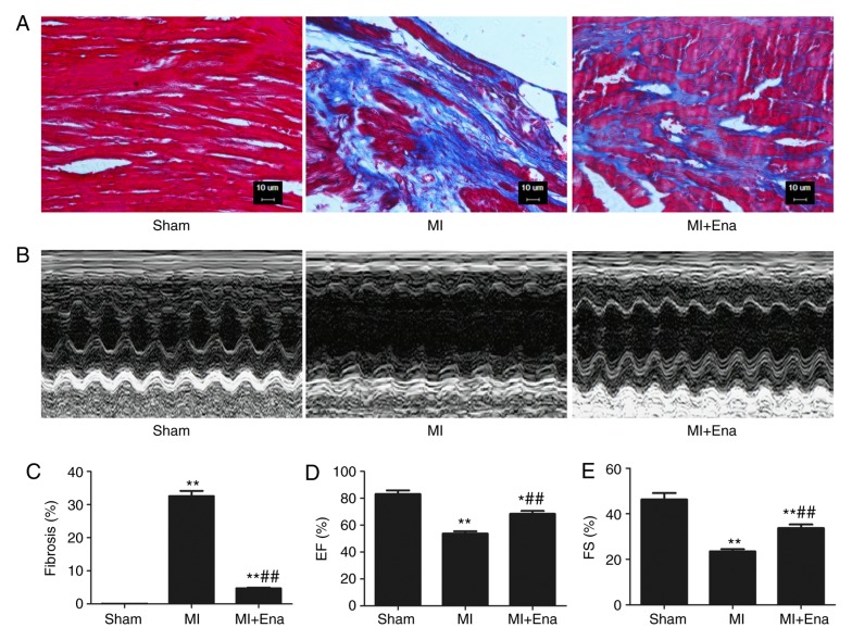
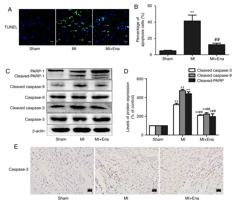
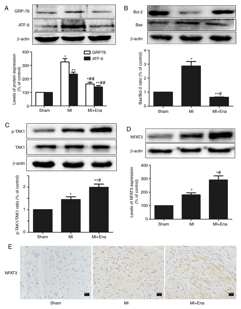
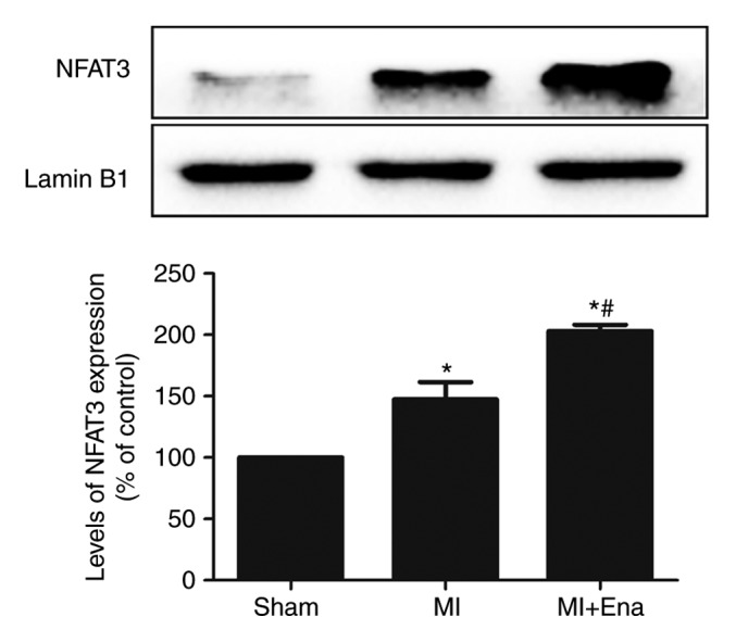
The present study investigated the effect of enalapril on myocardial infarction (MI) and its mechanism of action in mice. Treatment with enalapril significantly attenuated cellular apoptosis and death. In vivo, enalapril treatment alleviated MI injury, and decreased myocardial apoptosis and the size of the infarct area. This was paralleled by increased Bcl-2 expression, decreased Bax expression, a decreased caspase-3 level, decreased expression of endoplasmic reticulum stress-associated proteins, including activating transcription factor 6 and 78 kDa glucose-regulated protein, and fewer TUNEL-positive cells in the heart. Furthermore, enalapril-treatment increased transforming growth factor-activated kinase 1/nuclear factor of activated T cells 3 signaling, which protected the myocardium.

Keywords: apoptosis; cardiac remodeling; enalapril; myocardial infarction; transforming growth factor-β–activated kinase 1/nuclear factor of activated T cells pathway.


Figures

Figure 1.
Figure 1.
Enalapril decreases myocardial fibrosis and increases cardiac systolic function in the hearts of mice following MI. (A) Representative Masson's trichrome images of sections from the ischemic area of MI mice treated with enalapril or control. Scale bar, 10 µm. Magnification ×400. (B) Representative images produced by echocardiography from different groups of mice (sham, MI and MI treated with enalapril). (C) Fibrosis in sham, MI and MI+Ena groups. (D) The EF and (E) FS at the end of treatment with enalapril or control. *P<0.05 and **P<0.01 vs. sham group. ##P<0.01 vs. MI group. MI, myocardial infarction; EF, ejection fraction; FS, fraction shortening; Ena, enalapril.
Figure 2.
Figure 2.
Enalapril decreases myocardial apoptosis in MI mice. (A) Representative TUNEL immunofluorescence images in different groups of mice. The white arrows indicate the TUNEL-positive cells. Scale bar, 10 µm. Magnification, ×400. (B) Percentages of apoptosis-positive cells. (C) Endoplasmic reticulum stress-associated and mitochondrial dysfunction-associated proteins were detected by western blot analysis. Protein levels of cleaved PARP, cleaved caspase-3 and cleaved caspase-9 in the hearts of control mice, MI mice and MI mice treated with enalapril. (D) Optical density analysis of cleaved PARP, cleaved caspase-3 and cleaved caspase-9 in the heart tissues. (E) Immunohistochemical staining for cleaved caspase-3 in the hearts of control mice, MI mice and MI mice treated with enalapril. The data are presented as the mean ± standard deviation (n=6). *P<0.05 and **P<0.01 vs. sham group. ##P<0.01 vs. MI group. MI, myocardial infarction; PARP, poly (ADP-ribose) polymerase; Ena, enalapril.
Figure 3.
Figure 3.
Enalapril inhibits ER stress and mitochondrial dysfunction in MI mice. (A) Protein expression levels and quantitative analysis of GRP78 and ATF6. (B) Protein expression levels and quantitative analysis of Bcl-2 and Bax. (C) Protein expression levels and quantitative analysis of TAK1 and p-TAK1. (D) Protein expression levels and quantitative analysis of intracytoplasmic NFAT3. (E) Immunohistochemical staining for NFAT3 in the hearts of control mice, MI mice and MI mice treated with enalapril. Magnification, ×400. The data are presented as the mean ± standard deviation (n=6). *P<0.05 and **P<0.01 vs. sham group. #P<0.05 vs. the MI group. Ena, enalapril; ER, endoplasmic reticulum; MI, myocardial infarction; GRP78, 78 kDa glucose-regulated protein; ATF6, activating transcription factor 6; TAK1, transforming growth factor-β–activated kinase 1; p, phosphorylated; NFAT3, nuclear factor of activated T cells 3.
Figure 4.
Figure 4.
Nuclear protein expression levels and quantitative analysis of NFAT3 in MI mice. The data are presented as the mean ± standard deviation (n=6). *P<0.05 vs. sham group; #P<0.05 vs. MI group. Ena, enalapril; MI, myocardial infarction; NFAT3, nuclear factor of activated T cells 3.

Similar articles

Cited by

References

    1. Roth GA, Huffman MD, Moran AE, Feigin V, Mensah GA, Naghavi M, Murray CJ. Global and regional patterns in cardiovascular mortality from 1990 to 2013. Circulation. 2015;132:1667–1678. doi: 10.1161/CIRCULATIONAHA.114.008720. - DOI - PubMed
    1. Whelan RS, Kaplinskiy V, Kitsis RN. Cell death in the pathogenesis of heart disease: Mechanisms and significance. Annu Rev Physiol. 2010;72:19–44. doi: 10.1146/annurev.physiol.010908.163111. - DOI - PubMed
    1. Zhang T, Zhang Y, Cui M, Jin L, Wang Y, Lv F, Liu Y, Zheng W, Shang H, Zhang J, et al. CaMKII is a RIP3 substrate mediating ischemia- and oxidative stress-induced myocardial necroptosis. Nat Med. 2016;22:175–182. doi: 10.1038/nm.4017. - DOI - PubMed
    1. Messadi-Laribi E, Griol-Charhbili V, Gaies E, Vincent MP, Heudes D, Meneton P, Alhenc-Gelas F, Richer C. Cardioprotection and kallikrein-kinin system in acute myocardial ischaemia in mice. Clin Exp Pharmacol Physio. 2008;35:489–493. doi: 10.1111/j.1440-1681.2008.04902.x. - DOI - PubMed
    1. Investigators S, Yusuf S, Pitt B, Davis CE, Hood WB, Cohn JN. Effect of enalapril on survival in patients with reduced left ventricular ejection fractions and congestive heart failure. N Engl J Med. 1991;325:293–302. doi: 10.1056/NEJM199108013250501. - DOI - PubMed