Review
. 2020 Feb;19(2):1149-1154.
doi: 10.3892/etm.2019.8320. Epub 2019 Dec 12.

Current status of infarction in the basal ganglia-internal capsule due to mild head injury in children using PRISMA guidelines

Affiliations
Review

Current status of infarction in the basal ganglia-internal capsule due to mild head injury in children using PRISMA guidelines

Guangming Wang et al. Exp Ther Med. 2020 Feb.

Abstract

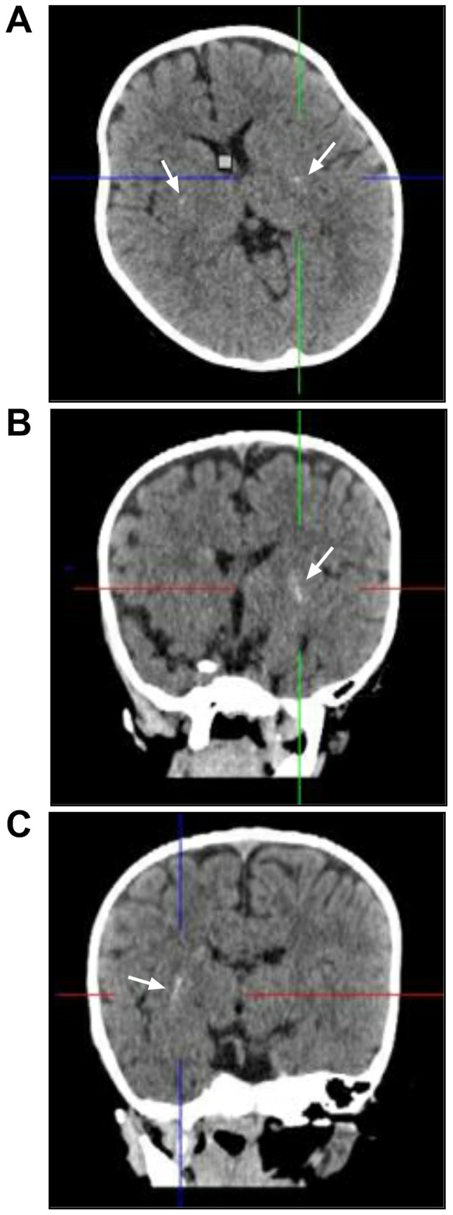
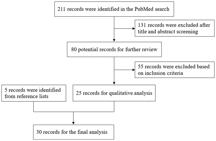
Post-traumatic basal ganglia-internal capsule (BGIC) infarction in pediatric patients is a relatively rare consequence of mild head injury (MHI). To the best of the authors' knowledge, at present, no comprehensive review has been published. To review research on BGIC infarction after MHI, a literature search was performed using the PubMed database and relevant search terms. According to recent data, MHI may cause BGIC infarction due to mechanical vasospasm of the perforating vessels in pediatric patients. The anatomical characteristics of the growing brain in infancy, mineralization of the lenticulostriate arteries and viral infection may all play a part in BGIC infarction after MHI, which often occurs within 24 months. Symptoms are not as severe and tend to disappear in the early period. Computed tomography or magnetic resonance imaging often shows BGIC infarction. There are also children with scattered calcification of the basal ganglia. Neural rehabilitation is a commonly accepted treatment. The prognosis of patients with BGIC infarction after MHI consistently improves.

Keywords: basal ganglia-internal capsule; children; infarction; mild head injury.


Figures

Figure 1.
Figure 1.
Computed tomography (CT) images of the illustrative case. (A) Head CT in axial view shows bilateral symmetric scattered mineralization of the lenticulostriate arteries (arrows). (B) Coronal CT shows scattered mineralization of the left lenticulostriate artery (arrow). (C) Coronal CT shows scattered mineralization of the right lenticulostriate artery (arrow). The brain tissue of this children is not well-developed, and the subarachnoid space is relatively large (A-C).
Figure 2.
Figure 2.
MRI of the illustrative case. (A) MRI T2 weighted image shows an infarct signal in the basal ganglia on either side (asterisks). (B) No abnormalities were found in the internal carotid artery system in the magnetic resonance angiogram. MRI, magnetic resonance imaging.
Figure 3.
Figure 3.
Flowchart showing how the present study was derived.
Figure 4.
Figure 4.
Mobile subarachnoid space segments of the lenticulostriate artery are more acute, shorter and more tensely stretched in (A) a child than in (B) an adult.

Similar articles

Cited by

References

    1. Nabika S, Kiya K, Satoh H, Mizoue T, Oshita J, Kondo H. Ischemia of the internal capsule due to mild head injury in a child. Pediatr Neurosurg. 2007;43:312–315. doi: 10.1159/000103313. - DOI - PubMed
    1. Kieslich M, Fiedler A, Heller C, Kreuz W, Jacobi G. Minor head injury as cause and co-factor in the aetiology of stroke in childhood: A report of eight cases. J Neurol Neurosurg Psychiatry. 2002;73:13–16. doi: 10.1136/jnnp.73.1.13. - DOI - PMC - PubMed
    1. Provenzale JM, Sorensen AG. Diffusion-weighted MR imaging in acute stroke: Theoretic considerations and clinical applications. AJR Am J Roentgenol. 1999;173:1459–1467. doi: 10.2214/ajr.173.6.10584783. - DOI - PubMed
    1. Jain P, Kishore P, Bhasin JS, Arya SC. Mineralizing angiopathy with basal ganglia stroke in an infant. Ann Indian Acad Neurol. 2015;18:233–234. doi: 10.4103/0972-2327.150619. - DOI - PMC - PubMed
    1. Yang FH, Wang H, Zhang JM, Liang HY. Clinical features and risk factors of cerebral infarction after mild head trauma under 18 months of age. Pediatr Neurol. 2013;48:220–226. doi: 10.1016/j.pediatrneurol.2012.12.008. - DOI - PubMed