. 2020 Feb;19(2):1225-1234.
doi: 10.3892/etm.2019.8349. Epub 2019 Dec 19.

Human cardiac extracellular matrix-chitosan-gelatin composite scaffold and its endothelialization

Affiliations

Human cardiac extracellular matrix-chitosan-gelatin composite scaffold and its endothelialization

Jingjing Lv et al. Exp Ther Med. 2020 Feb.

Abstract

The present study developed a cardiac extracellular matrix-chitosan-gelatin (cECM-CG) composite scaffold that can be used as a tissue-engineered heart patch and investigated its endothelialization potential by incorporating CD34+ endothelial progenitor cells (EPCs). The cECM-CG composite scaffold was prepared by blending cardiac extracellular matrix (cECM) with biodegradable chitosan-gelatin (CG). The mixture was lyophilized using vacuum freeze-drying. CD34+ EPCs were isolated and seeded on the scaffolds, and then the endothelialization effect was subsequently investigated. Effects of the scaffolds on CD34+ EPCs survival and proliferation were evaluated by immunofluorescence staining and MTT assay. Cell differentiation into endothelial cells and the influence of the scaffolds on cell differentiation were investigated by reverse transcription-quantitative PCR (RT-qPCR), immunofluorescence staining and tube formation assay. The present results indicated that most cells were removed after decellularization, but the main extracellular matrix components were retained. Scanning electron microscopy imaging illustrated three-dimensional and porous scaffolds. The present results suggested the cECM-CG composite scaffold had a higher water absorption ability compared with the CG scaffold. Additionally, compared with the CG scaffold, the cECM-CG composite scaffold significantly increased cell survival and proliferation, which suggested its non-toxicity and biocompatibility. Furthermore, RT-qPCR, immunofluorescence and tube formation assay results indicated that CD34+ EPCs differentiated into endothelial cells, and the cECM-CG composite scaffold promoted this differentiation process. In conclusion, the present results indicated that the human cECM-CG composite scaffold generated in the present study was a highly porous, biodegradable three-dimensional scaffold which supported endothelialization of seeded CD34+ EPCs. The present results suggested that this cECM-CG composite scaffold may be a promising heart patch for use in heart tissue engineering for congenital heart disease.

Keywords: cardiac extracellular matrix; endothelial progenitor cells; endothelialization; heart patch; scaffold.


Figures

Figure 1.
Figure 1.
Preparation and characterization of cECM and CG/cECM-CG scaffolds. Comparison of cECM composition of heart tissue before and after decellularization. (A) Hematoxylin and eosin staining was performed to confirm complete decellularization. Scale bar, 10 µm. (B) Masson staining was performed for collagen fiber evaluation, collagen fibers were stained blue. Scale bar, 50 µm. (C) Van Gieson staining was performed for elastic fiber evaluation, elastic fibers were stained yellow. Scale bar, 50 µm. Immunohistochemical staining of (D) fibronectin and (E) laminin, fibronectin and laminin were stained brown. Scale bar, 50 µm. (F) Statistical analysis of DNA content. n=3 in each group. (G) Statistical analysis of GAG content of heart tissue before and after decellularization. (H) Macrostructure of the scaffold. SEM analysis of (I) CG and (J) cECM-CG composite scaffolds. (K) Evaluation of the water absorption capacity of the scaffolds. GAG, glycosaminoglycans; cECM, cardiac extracellular matrix; CG, chitosan-gelatin; cECM-CG, cardiac extracellular matrix-chitosan-gelatin.
Figure 2.
Figure 2.
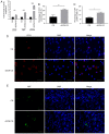
Scaffold composited with cECM improves cell survival and promotes proliferation of CD34+ EPCs. (A) The population of the sorted cells. (B) The purity of CD34+ EPCs after magnetic cell sorting isolation. (C) Cell viability assay analyzed the viability of CD34+ EPCs seeded on the scaffolds at day 5 by fluorescence microscopy. Living cells were stained green by calcein-AM. Dead cells were stained red by propidium iodide. Scale bar, 20 µm. (D) Representative images of cell proliferation assessed by Ki67 immunofluorescence staining at day 10. Positive cells were stained green. Scale bar, 100 µm. (E) Proliferation was analyzed by MTT assay on days 1, 3, 5, 7, 10 and 14. n=3 in each group. (F) Percentage of Ki67 positive cells using immunofluorescence staining was calculated by counting the positive cells in three different and randomly chosen view fields. **P<0.01 vs. CG. cECM, cardiac extracellular matrix; CG, chitosan-gelatin; cECM-CG, cardiac extracellular matrix-chitosan-gelatin; EPC, endothelial progenitor cells.
Figure 3.
Figure 3.
In vitro differentiation of CD34+ EPCs into endothelial cells cultured on CG and cECM-CG composite scaffolds. (A) Reverse transcription-quantitative PCR results showed that the CD34+ EPCs seeded on cECM-CG scaffold upregulated the gene expression levels of EC markers including CD31, vWF and CD144 on day 21, compared with cells seeded on CG scaffold. Percentages of (B) CD31-positive and (C) vWF-positive cells were calculated at day 21 in three different and randomly chosen view fields. CD34+ EPCs on cECM-CG scaffold showed a higher differentiation rate compared with CG. The experiment was repeated three times. Representative images of immunofluorescence staining of the expression levels of (D) CD31 and (E) vWF. Scale bar, 50 µm. *P<0.05, **P<0.01, ***P<0.001 vs. CG. cECM, cardiac extracellular matrix; CG, chitosan-gelatin; cECM-CG, cardiac extracellular matrix-chitosan-gelatin; EPC, endothelial progenitor cells; vWF, von Willebrand factor.
Figure 4.
Figure 4.
cECM-CG composite scaffold-based conditioned medium increases tube formation of HUVECs. (A and B) Representative images of the tube formation capacity of HUVECs induced by conditioned medium from CD34+ cells cultured on (A) CG and (B) cECM-CG scaffolds. Scale bar, 100 µm. Quantitative analysis of the (C) branch points and (D) tube length of both groups. *P<0.05 vs. CG. CG, chitosan-gelatin; cECM-CG, cardiac extracellular matrix-chitosan-gelatin; EPC, endothelial progenitor cells; vWF, von Willebrand factor; HUVECs, human umbilical vein endothelial cells.

Similar articles

Cited by

References

    1. Benjamin EJ, Muntner P, Alonso A, Bittencourt MS, Callaway CW, Carson AP, Chamberlain AM, Chang AR, Cheng S, Das SR, et al. Heart disease and stroke statistics-2019 update: A report from the American heart association. Circulation. 2019;139:e56–e528. doi: 10.1161/CIR.0000000000000659. - DOI - PubMed
    1. Tsilimigras DI, Oikonomou EK, Moris D, Schizas D, Economopoulos KP, Mylonas KS. Stem cell therapy for congenital heart disease: A systematic review. Circulation. 2017;136:2373–2385. doi: 10.1161/CIRCULATIONAHA.117.029607. - DOI - PubMed
    1. Kochupura PV, Azeloglu EU, Kelly DJ, Doronin SV, Badylak SF, Krukenkamp IB, Cohen IS, Gaudette GR. Tissue-engineered myocardial patch derived from extracellular matrix provides regional mechanical function. Circulation. 2005;112(Suppl 9):I144–I149. - PubMed
    1. Mosala Nezhad Z, Poncelet A, Fervaille C, Gianello P. Comparing the host reaction to CorMatrix and different cardiac patch materials implanted subcutaneously in Growing Pigs. Thoracic Cardiovasc Surg. 2019;67:44–49. doi: 10.1055/s-0037-1607332. - DOI - PubMed
    1. Nishida H, Nakatsuka D, Kawano Y, Hiraiwa N, Takanashi S, Tabata M. Outcomes of totally endoscopic atrial septal defect closure using a glutaraldehyde-treated autologous pericardial patch. Circ J. 2017;81:689–693. doi: 10.1253/circj.CJ-16-0888. - DOI - PubMed