. 2020 Feb 1;11(1):1-16.
doi: 10.14336/AD.2019.0507. eCollection 2020 Feb.

Neuropsychological Deficits Chronically Developed after Focal Ischemic Stroke and Beneficial Effects of Pharmacological Hypothermia in the Mouse

Affiliations

Neuropsychological Deficits Chronically Developed after Focal Ischemic Stroke and Beneficial Effects of Pharmacological Hypothermia in the Mouse

Weiwei Zhong et al. Aging Dis. .

Abstract

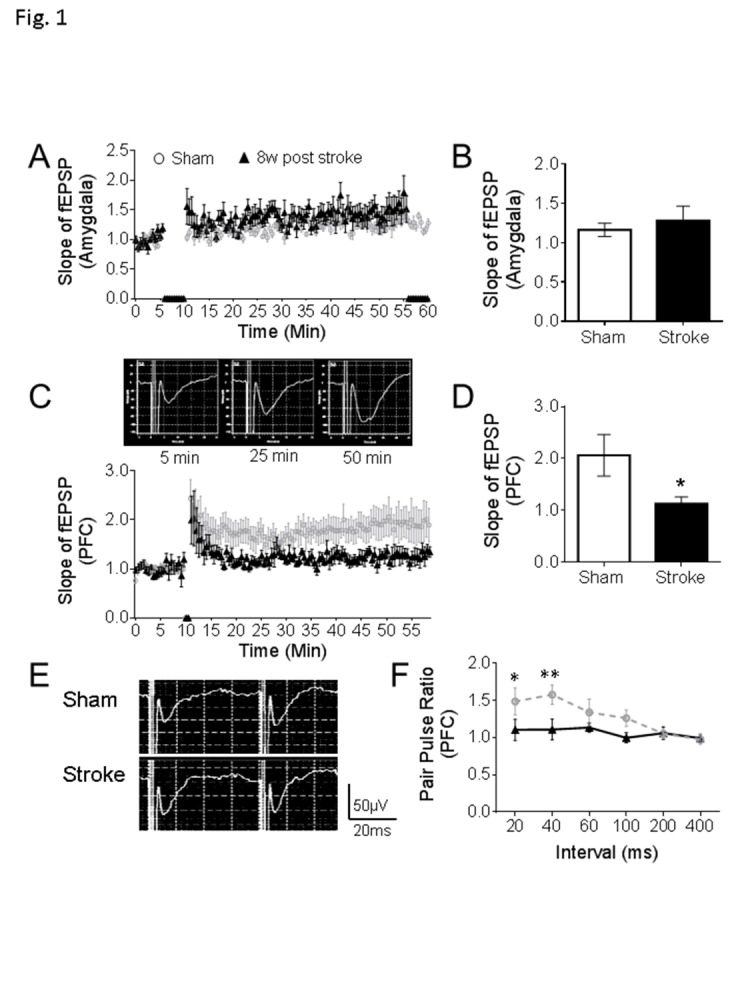
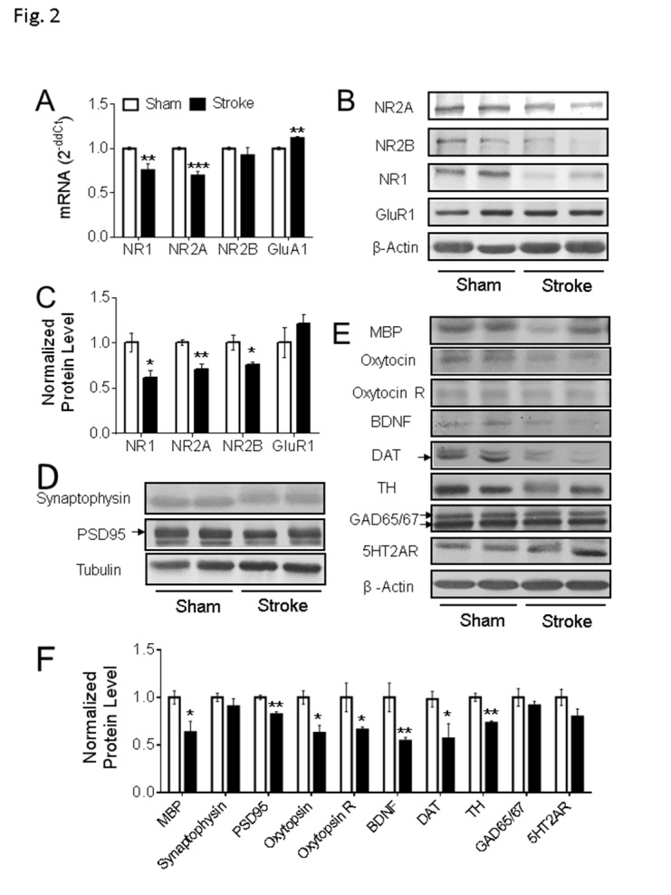
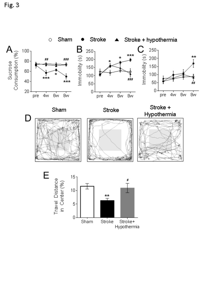
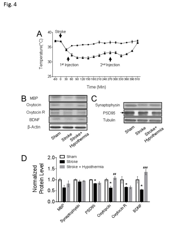
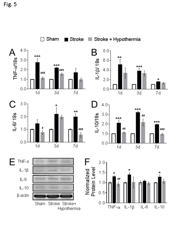
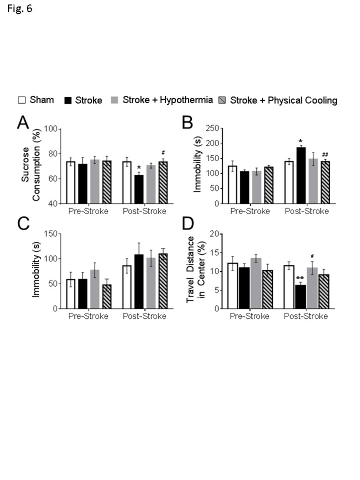
Stroke is a leading cause of human death and disability, with around 30% of stroke patients develop neuropsychological/neuropsychiatric symptoms, such as post-stroke depression (PSD). Basic and translational research on post-stroke psychological disorders is limited. In a focal ischemic stroke mouse model with selective damage to the sensorimotor cortex, sensorimotor deficits develop soon after stroke and spontaneous recovery is observed in 2-4 weeks. We identified that mice subjected to a focal ischemic insult gradually developed depression/anxiety like behaviors 4 to 8 weeks after stroke. Psychological/psychiatric disorders were revealed in multiple behavioral examinations, including the forced swim, tail suspension, sucrose preference, and open field tests. Altered neuronal plasticity such as suppressed long-term potentiation (LTP), reduced BDNF and oxytocin signaling, and disturbed dopamine synthesis/uptake were detected in the prefrontal cortex (PFC) during the chronic phase after stroke. Pharmacological hypothermia induced by the neurotensin receptor 1 (NTR1) agonist HPI-363 was applied as an acute treatment after stroke. A six-hr hypothermia treatment applied 45 min after stroke prevented depression and anxiety like behaviors examined at 6 weeks after stroke, as well as restored BDNF expression and oxytocin signaling. Additionally, hypothermia induced by physical cooling also showed an anti-depression and anti-anxiety effect. The data suggested a delayed beneficial effect of acute hypothermia treatment on chronically developed post-stroke neuropsychological disorders, associated with regulation of synaptic plasticity, neurotrophic factors, dopaminergic activity, and oxytocin signaling in the PFC.

Keywords: BDNF; oxytocin; pharmacological hypothermia; post-stroke depression (PSD); stroke.


Conflict of interest statement

Conflict of interest The authors declare no conflicts of interest.

Figures

Figure 1.
Figure 1.
Defects of synaptic plasticity in the prefrontal cortex after focal ischemic stroke. (A-B) In the amygdala, stable fEPSPs were recorded for 6 min as a baseline, high frequency stimulation (HFS) induced an LTP-like response, but no significant differences were found between stroke mice and sham (sham: n = 8, stroke: n = 9). (C-D) In the PFC of sham mice, HFS initiated the LTP as displayed in the representative traces. In the comparison to sham animals, smaller LTPs were recorded in the PFC of mice at 8 weeks after stroke (C). At 50 min of the recording, the slope of fEPSP was significantly reduced in the PFC of stroke mice than sham controls (D). (E-F) Representative traces are pair-pulse response with a 40 ms interval in PFC. Stroke insult notably reduced the pair-pulse ratio of fEPSPs at 20 ms and 40 ms intervals (Sham: n = 8, Stroke: n = 9; *P < 0.05, **P < 0.01; Student’s t-test).
Figure 2.
Figure 2.
Gene expression in the PFC of focal ischemic stroke mice. (A-C) In the ipsilateral PFC of the animals with stroke for 8 weeks, qPCR data suggested the mRNA level of NR1 and NR2A were significantly reduced (A, Sham: n = 5, Stroke: n = 5), which is consistent with the western blot results (B-C, Sham: n = 4, Stroke: n = 4). The mRNA level of GluR1 was significantly increased (A), although no significant difference was detected in the protein expression level of GluR1 (B-C) (*P < 0.05, **P < 0.01, ***P < 0.001; Student’s t-test). (D-F) In the western blot assessment, compared to the animals in the sham group, the protein expression levels of MBP, PSD95, oxytocin, oxytocin R, BDNF, DAT, and TH were significantly reduced in PFC of stroke animals. Arrows point to the bands of PSD95 and DAT; the two GAD65/67 bands were used in the quantification. (Sham: n = 4, Stroke: n = 5; *P < 0.05, **P < 0.01; Student’s t-test).
Figure 3.
Figure 3.
Delayed development of neuropsychological behaviors and beneficial effects of pharmacological hypothermia after focal ischemic stroke. (A) The sucrose preference test was used to test anhedonic behavior in mice. In comparison to the sham, the stroke animals consumed significantly less sucrose 2-8 weeks post stroke and acute hypothermia treatment significantly increased the sucrose consumption at 4 weeks and 8 weeks post stroke. The main factor of treatment showed a significant difference (F=16.3, df =2, P < 0.001). No significant differences were found in the main factor of time (F = 2.24, df = 3, P = 0.088) or the interactions of time × treatment (F = 1.90, df = 6, P = 0.088). (B-C) The tail suspension (B) and forced swim test (C) were used to test the depression like behavior in mice. (B) In the tail suspension test, the stroke mice showed significantly increased immobile durations at 4-8 weeks after stroke and acute hypothermia treatment significantly reduced the immobility at 6 weeks and 8 weeks after stroke. Significant differences were detected in the main factor of time (F = 6.01, df = 3, P < 0.001), treatment (F = 11.24, df = 2, P < 0.001) and interaction of time × treatment (F = 4.79, df = 6, P < 0.001). (C) In the forced swim test, the stroke mice were immobile significantly longer than the sham and acute hypothermia treatment significantly reduced the immobility in stroke mice. There are significant differences in the main factor of time (F = 3.90, df = 3, P < 0.05) and treatment (F = 3.60, df = 2, P < 0.05). No significant difference was found in the interaction of time × treatment (F = 1.55, df = 6, P = 0.172) (D-E) The open field test was used to test the anxious-like behavior in mice. The walking traces of the test mice in the open field were shown as a representative (D). The animals traveled significantly less in the central region of the arena than sham at 6-7 weeks after stroke, while acute hypothermia treatment increased the ratio of traveling in the center region in these mice (E). (A-E, sham: n = 10, stroke: n = 10, stroke + hypothermia: n = 8) (* sham vs stroke; # stroke vs stroke + hypothermia; *, # P < 0.05, **, ## P < 0.01, ***, ### P < 0.001; Two-way ANOVA and One-way ANOVA with Fisher’s post hoc)
Figure 4.
Figure 4.
Chronic consequences in the non-ischemic PFC after acute pharmacological hypothermia in focal ischemic stroke mice. (A) The time course of pharmacologically induced hypothermia with HPI363 in mice after stroke (stroke: n = 10, stroke + hypothermia: n = 8). (B-D) The western blot results showed both the expression levels of oxytocin and BDNF were significantly increased in the stroke mice with hypothermia treatment. Only the active form of BDNF band and the arrow pointed PSD95 band were used in the quantification. (sham: n = 4, stroke: n = 5, stroke + hypothermia: n = 4) (* sham vs stroke; # stroke vs stroke + hypothermia; * P < 0.05, **, ## P < 0.01, ***, ### P < 0.001; One-way ANOVA with Fisher’s post hoc)
Figure 5.
Figure 5.
Acute hypothermia treatment altered the cytokines expression in the non-ischemic PFC. (A-D) The mRNA levels of cytokines were measured using qPCR analyses in the ipsilateral PFC at 1day, 3days and 7days after stroke. Stroke induction significantly enhanced the mRNAs of the pro-inflammatory cytokines TNF-α (A), IL-1β (B) and IL-6 (C) at 1day, 3day, and 7days post stroke. Hypothermia treatment significantly attenuated the TNF-α (A) at 1day and 3days post stroke and IL-6 at 1day and 7days post stroke. On the contrary, the mRNA of anti-inflammatory factor IL-10 (D) was significantly increased in stroke mice and hypothermia treatment reduced the IL-10 mRNA level in stroke mice at 1 day, 3 day and 7 days post-surgery. (n = 3 in each groups) (E-F) Western blot was used to quantify the cytokine levels in PFC at 3 days post stroke. Stroke induction enhanced the protein expression of TNF-α, IL-1β, and IL-10, while acute hypothermia treatment significantly reduced the TNF-α expression level in PFC at 3 days post stroke. No change was observed in the pro-inflammation cytokine IL-6 between groups. Only the active form of IL-1β was used in the quantification. (sham: n = 4, stroke: n = 4, stroke + hypothermia: n = 4) (* sham vs stroke; # stroke vs stroke + hypothermia; *, # P < 0.05, **, ## P < 0.01, ***, ### P < 0.001; One-way ANOVA with Fisher’s post hoc)
Figure 6.
Figure 6.
Physical cooling alleviated psychological behaviors in mice after stroke. The behavioral assessments were performed at 6-7 weeks post stroke in the experimental mice. (A) In the sucrose preference test, physical cooling prevented the loss of sucrose preference to levels similar to the drug induced hypothermia group. (B) In the tail suspension test, physical cooling significantly attenuated the immobility in stroke mice. (C) No significant differences were detected between groups in the forced swim test. (D) In the open field test, although physical cooling failed to significantly increase the travel distance ratio in the center, no differences were found between sham and stroke mice with physical cooling treatment. (sham: n = 10, stroke: n = 10, stroke + hypothermia: n = 8, stroke + physical cooling: n = 8; * sham vs stroke; # stroke vs stroke + hypothermia; *, # P < 0.05, **, ## P < 0.01; One-way ANOVA with Fisher’s post hoc)

Similar articles

Cited by

References

    1. Folstein MF, Maiberger R, McHugh PR (1977). Mood disorder as a specific complication of stroke. J Neurol Neurosurg Psychiatry, 40: 1018-1020 - PMC - PubMed
    1. Chun HY, Whiteley WN, Dennis MS, Mead GE, Carson AJ (2018). Anxiety After Stroke: The Importance of Subtyping. Stroke, 49: 556-564 - PMC - PubMed
    1. Ayerbe L, Ayis S, Wolfe CD, Rudd AG (2013). Natural history, predictors and outcomes of depression after stroke: systematic review and meta-analysis. Br J Psychiatry, 202: 14-21 - PubMed
    1. Kim JS (2016). Post-stroke Mood and Emotional Disturbances: Pharmacological Therapy Based on Mechanisms. J Stroke, 18: 244-255 - PMC - PubMed
    1. Kronenberg G, Balkaya M, Prinz V, Gertz K, Ji S, Kirste I, et al. (2012). Exofocal dopaminergic degeneration as antidepressant target in mouse model of poststroke depression. Biol Psychiatry, 72: 273-281 - PubMed

LinkOut - more resources