Review
. 2020 Feb 1;11(1):200-211.
doi: 10.14336/AD.2020.0103. eCollection 2020 Feb.

The Interstitial System of the Brain in Health and Disease

Affiliations
Review

The Interstitial System of the Brain in Health and Disease

Ashok K Shetty et al. Aging Dis. .

Abstract

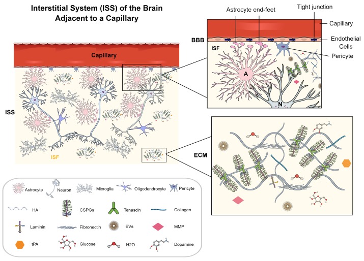
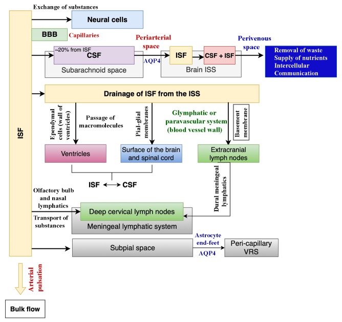
The brain interstitial fluid (ISF) and the cerebrospinal fluid (CSF) cushion and support the brain cells. The ISF occupies the brain interstitial system (ISS), whereas the CSF fills the brain ventricles and the subarachnoid space. The brain ISS is an asymmetrical, tortuous, and exceptionally confined space between neural cells and the brain microvasculature. Recently, with a newly developed in vivo measuring technique, a series of discoveries have been made in the brain ISS and the drainage of ISF. The goal of this review is to confer recent advances in our understanding of the brain ISS, including its structure, function, and the various processes mediating or disrupting ISF drainage in physiological and pathological conditions. The brain ISF in the deep brain regions has recently been demonstrated to drain in a compartmentalized ISS instead of a highly connected system, together with the drainage of ISF into the cerebrospinal fluid (CSF) at the surface of the cerebral cortex and the transportation from CSF into cervical lymph nodes. Besides, accumulation of tau in the brain ISS in conditions such as Alzheimer's disease and its link to the sleep-wake cycle and sleep deprivation, clearance of ISF in a deep sleep via increased CSF flow, novel approaches to remove beta-amyloid from the brain ISS, and obstruction to the ISF drainage in neurological conditions are deliberated. Moreover, the role of ISS in the passage of extracellular vesicles (EVs) released from neural cells and the rapid targeting of therapeutic EVs into neural cells in the entire brain following an intranasal administration, and the promise and limitations of ISS based drug delivery approaches are discussed.

Keywords: beta-amyloid; cerebrospinal fluid; extracellular matrix; extracellular vesicles; glymphatic system; interstitial fluid; phosphorylated tau.


Figures

Figure 1.
Figure 1.
A cartoon illustrating the brain interstitial system (ISS) between neural cells comprising interstitial fluid (ISF) and the extracellular matrix (ECM), adjacent to a brain microvasculature. The magnified view of a portion of the cartoon on the top right shows endothelial cells with tight junctions, astrocyte end-feet, and pericytes at the interface of ISS and the microvasculature. The magnified view on the bottom right shows the ECM with its structure and components. The ECM contains hyaluronic acid, proteoglycans - CSPGs, tenascin, and a small amount of collagen, laminin, and fibronectin. Some components of ISF are represented, including EVs, H2O, glucose, dopamine, matrix metallopeptidase (MMP), and tissue plasminogen activators (tPAs). The illustrated ECM is distributed as the neural interstitial matrix in the space between neural cells. A, astrocyte; BBB, blood-brain barrier; N, neuron.
Figure 2.
Figure 2.
A schematic showing the various communications of the interstitial fluid (ISF), and the mechanisms by which ISF is drained into the lymphatic system. ISF interacts with neurons through the capillaries, and the substance exchange between capillaries and neurons is facilitated by the blood-brain barrier (BBB). The cerebrospinal fluid (CSF) resides in the subarachnoid space, and ~20% of the CSF in the brain comes from the ISF. The CSF influx into the brain happens through the periarterial spaces, the convective flow into the ISS occurs through the water channel aquaporin-4 (AQP4, located on astrocyte end-feet) where CSF mixes with the ISF, and then the efflux of ISF occurs through perivenous spaces. The drainage of ISF occurs through three suggested conduits. These include passage through ependymal cells into ventricles, pia-glial membranes into the surface of the brain and spinal cord, and the glymphatic system into extracranial lymph nodes. The ISF is then transported via dural meningeal lymphatics into the deep cervical lymph nodes. Also, the transportation of substances from the ISF into the deep cervical lymph nodes can occur via the olfactory bulb and nasal lymphatics. Furthermore, the end-feet of astrocytes are also involved in the transportation of ISF from the subpial space into the peri-capillary Virchow-Robin space (VRS) through AQP4 channels. The arterial pulsation promotes the ISF bulk flow.

References

    1. Lei Y, Han H, Yuan F, Javeed A, Zhao Y (2017). The brain interstitial system: Anatomy, modeling, in vivo measurement, and applications. Prog Neurobiol, 157:230-246. - PubMed
    1. Chatterjee K, Esparza CM, Munson JM (2019). Methods to measure, model and manipulate fluid flow in brain. J Neurosci Methods: 108541. - PMC - PubMed
    1. Pajevic S (2019). Exploring the Dynamics of Brain Extracellular Space. Biophys J, 117):1781-1782. - PMC - PubMed
    1. Nicholson C, Kamali-Zare P, Tao L (2011). Brain Extracellular Space as a Diffusion Barrier. Comput Vis Sci, 14:309-325. - PMC - PubMed
    1. Bielas S, Higginbotham H, Koizumi H, Tanaka T, Gleeson JG (2004). Cortical neuronal migration mutants suggest separate but intersecting pathways. Annu Rev Cell Dev Biol, 20:593-618. - PubMed

LinkOut - more resources