. 2019 Dec 6;11(1):99-108.
doi: 10.1364/BOE.11.000099. eCollection 2020 Jan 1.

Multiplex protein-specific microscopy with ultraviolet surface excitation

Affiliations

Multiplex protein-specific microscopy with ultraviolet surface excitation

Jiaming Guo et al. Biomed Opt Express. .

Abstract

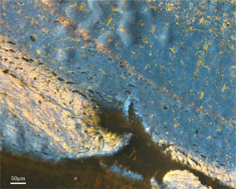
Immunohistochemical techniques, such as immunofluorescence (IF) staining, enable microscopic imaging of local protein expression within tissue samples. Molecular profiling enabled by IF is critical to understanding pathogenesis and is often involved in complex diagnoses. A recent innovation, known as microscopy with ultraviolet surface excitation (MUSE), uses deep ultraviolet (≈280 nm) illumination to excite labels at the tissue surface, providing equivalent images without fixation, embedding, and sectioning. However, MUSE has not yet been integrated into traditional IF pipelines. This limits its application in more complex diagnoses that rely on protein-specific markers. This paper aims to broaden the applicability of MUSE to multiplex immunohistochemistry using quantum dot nanoparticles. We demonstrate the advantages of quantum dot labels for protein-specific MUSE imaging on both paraffin-embedded and intact tissue, significantly expanding MUSE applicability to protein-specific applications. Furthermore, with recent innovations in three-dimensional ultraviolet fluorescence microscopy, this opens the door to three-dimensional IF imaging with quantum dots using ultraviolet excitation.


Conflict of interest statement

The authors declare that there are no conflicts of interest related to this article.

Figures

Fig. 1.
Fig. 1.
Properties of ThermoFisher QD streptavidin conjugates. (a) Using indirect IHC, the primary antibody (blue) binds the antigen and the QD-tagged secondary antibody (orange) binds to the primary. A close-up showing the structural properties of a QD streptavidin conjugate. (b) Absorption (dashed curve) and emission (solid curve) spectra of several QDs showing capability of multiplex mapping with a single deep UV excitation band. An average excitation spectrum of all QDs is plotted (black dashed curve). QDs in other sizes (ex. 525 nm, 625 nm, 705 nm) are also commercially available.
Fig. 2.
Fig. 2.
Concept of MUSE imaging. (a) Side view of the MUSE construct showing (1) a sample stage, (2) a UV LED, and (3) a standard microscope objective. A close-up view of the imaging region showing the reduced UV penetration depth in tissue. (b) An RGB image of the cut surface of a 1 mm thick mouse brain coronal section perfused with fluorescent tattoo ink and labelled with QD 585 for NeuN-positive neurons.
Fig. 3.
Fig. 3.
Examining QDs with deep-UV excitation. GFAP-positive cells were labelled with different QDs (left to right) that emit respectively at 565 nm, 585 nm, 605 nm, and 655 nm and imaged with a wide-field fluorescence microscope (top) and MUSE (bottom).
Fig. 4.
Fig. 4.
Protein-specific triplex MUSE imaging of a coronal paraffin-embedded mouse brain section, in which NeuN-positive neurons (yellow) were labeled with QD 585, CollIV-positive vessels (orange-brown) were labeled with QD 605, and GFAP-positive astrocytes (bright red-pink) were labeled with QD 655. Different tissue regions such as the hippocampal formation (denoted as HPF) and thalamus (denoted as TH) are visible under UV excitation, and individual cell groups such as medial habenula (denoted as MH) and lateral habenula (denoted as LH) are also distinguishable in the epithalamus areas. Anatomical details such corpus callosum (denoted as CC) and myelinated fiber tracts are resolved and rendered in blue via tissue autofluorescence under UV light.
Fig. 5.
Fig. 5.
Protein-specific triplex MUSE imaging of a coronal paraffin-embedded mouse brain section, in which GFAP-positive astrocytes were labeled with QD 585, MBP-positive myelin was labeled with QD 605, and NeuN-positive neurons were stained with QD 655 in the hippocampus. Colors were manually edited to enhance image contrast.
Fig. 6.
Fig. 6.
Coronal MUSE imaging of a thick intact brain section stained with three different types of stains. GFAP-positive astrocytes (denoted as A) were labeled with QD 585, all cell nuclei (denoted as C) were counter-stained with HO342, and vessels (denoted as V) were stained with India ink during lumen perfusion.

Similar articles

Cited by

References

    1. Hurley M. J., Brandon B., Gentleman S. M., Dexter D. T., “Parkinson’s disease is associated with altered expression of CaV1 channels and calcium-binding proteins,” Brain 136(7), 2077–2097 (2013).10.1093/brain/awt134 - DOI - PubMed
    1. Castillo-Carranza D. L., Nilson A. N., Van Skike C. E., Jahrling J. B., Patel K., Garach P., Gerson J. E., Sengupta U., Abisambra J., Nelson P., Troncoso J., Ungvari Z., Galvan V., Kayed R., “Cerebral microvascular accumulation of tau oligomers in alzheimer’s disease and related tauopathies,” Aging Dis. 8(3), 257–266 (2017).10.14336/AD.2017.0112 - DOI - PMC - PubMed
    1. Fereidouni F., Harmany Z. T., Tian M., Todd A., Kintner J. A., McPherson J. D., Borowsky A. D., Bishop J., Lechpammer M., Demos S. G., Levenson R., “Microscopy with ultraviolet surface excitation for rapid slide-free histology,” Nat. Biomed. Eng. 1(12), 957–966 (2017).10.1038/s41551-017-0165-y - DOI - PMC - PubMed
    1. Xie W., Chen Y., Wang Y., Wei L., Yin C., Glaser A. K., Fauver M. E., Seibel E. J., Dintzis S. M., Vaughan J. C., Reder N. P., Liu J. T. C., “Microscopy with ultraviolet surface excitation for wide-area pathology of breast surgical margins,” J. Biomed. Opt. 24(02), 1 (2019).10.1117/1.JBO.24.2.026501 - DOI - PMC - PubMed
    1. Duraiyan J., Govindarajan R., Kaliyappan K., Palanisamy M., “Applications of immunohistochemistry,” J. Pharm. BioAllied Sci. 4(6), 307–S309 (2012).10.4103/0975-7406.100281 - DOI - PMC - PubMed

LinkOut - more resources