. 2020 Jan 17;6(3):eaaw9727.
doi: 10.1126/sciadv.aaw9727. eCollection 2020 Jan.

When and how self-cleaning of superhydrophobic surfaces works

Affiliations

When and how self-cleaning of superhydrophobic surfaces works

Florian Geyer et al. Sci Adv. .

Abstract

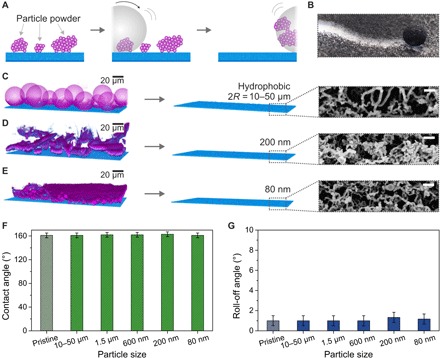
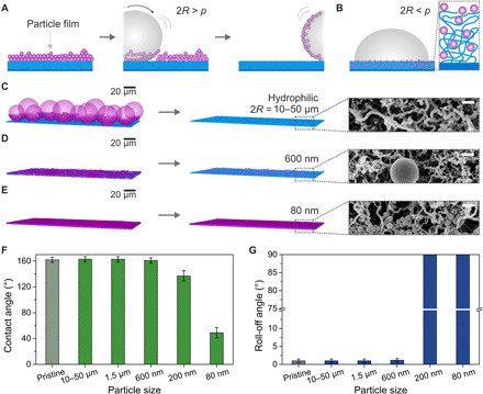
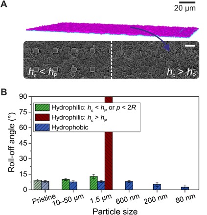
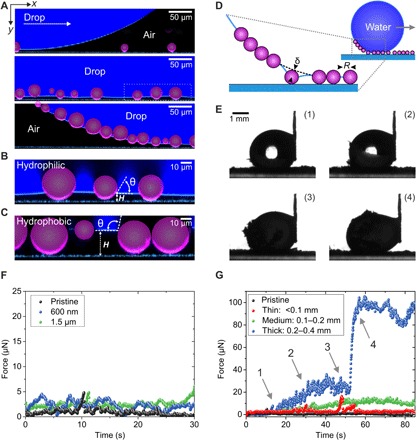
Despite the enormous interest in superhydrophobicity for self-cleaning, a clear picture of contaminant removal is missing, in particular, on a single-particle level. Here, we monitor the removal of individual contaminant particles on the micrometer scale by confocal microscopy. We correlate this space- and time-resolved information with measurements of the friction force. The balance of capillary and adhesion force between the drop and the contamination on the substrate determines the friction force of drops during self-cleaning. These friction forces are in the range of micro-Newtons. We show that hydrophilic and hydrophobic particles hardly influence superhydrophobicity provided that the particle size exceeds the pore size or the thickness of the contamination falls below the height of the protrusions. These detailed insights into self-cleaning allow the rational design of superhydrophobic surfaces that resist contamination as demonstrated by outdoor environmental (>200 days) and industrial standardized contamination experiments.


Figures

Fig. 1
Fig. 1. Self-cleaning of superhydrophobic surfaces.
(A) The surfaces are contaminated with particles of different sizes (80 nm to 50 μm) and polarities (hydrophobic/hydrophilic). (B) Water drops roll over the contaminated surface. (C) Can the water drops remove the contamination and how is superhydrophobicity affected? How does the self-cleaning evolve on the micrometer scale, and which forces are involved in the self-cleaning process?
Fig. 2
Fig. 2. Effect of differently sized hydrophobic particle powder contamination on nanoporous superhydrophobic surfaces.
(A) Schematic illustration of the self-cleaning process of hydrophobic particle powders (purple) by a water drop (gray) on a superhydrophobic surface (blue). Colors and texture were chosen to match the LSCM images. (B) Photograph of a 10-μl water drop cleaning a nanoporous superhydrophobic surface contaminated with Oil Red dye particles (appear black). (C to E) LSCM images after contaminating the nanoporous superhydrophobic surface (left) with powders of hydrophobic particles with diameters of 10 to 50 μm, 200 nm, and 80 nm (see Materials and Methods for details of the image processing). Efficient cleaning of all hydrophobic powders was verified by LSCM (center) and SEM (right). Scale bars, 200 nm (SEM). (F and G) Contact and roll-off angles using 6-μl water drops after self-cleaning of a nanoporous surface consecutively contaminated with hydrophobic particle powders.
Fig. 3
Fig. 3. Effect of hydrophilic particle contamination having various particle sizes deposited from ethanol dispersion on nanoporous superhydrophobic surfaces.
(A) Schematic illustration of the self-cleaning process of hydrophilic particles (purple; 2R > p) deposited from ethanol dispersion by a water drop (gray). (B) Particles of smaller diameter than the pore diameter (2R < p) can penetrate the coating (blue), affecting wetting properties. (C to E) LSCM images (left) after contamination of the superhydrophobic surfaces with hydrophilic particles with diameters of 10 to 50 μm, 600 nm, and 80 nm (see Materials and Methods and fig. S9 for details of image processing). LSCM (center) and SEM images (right) show the surfaces after rinsing. Scale bars, 200 nm (SEM). (F and G) Contact and roll-off angles using 6-μl water drops after self-cleaning of nanoporous surfaces contaminated with various hydrophilic particles (dried from ethanol dispersion).
Fig. 4
Fig. 4. Contamination and self-cleaning of superhydrophobic microstructured SU-8 pillars (rectangular, 10-μm height with 5 × 5–μm2 top areas; center-center distance of pillars, 20 μm).
(A) LSCM (top) and SEM (bottom) images showing a surface contaminated with 1.5-μm particles. On the left side of the SEM image, the micropillar array is only partially filled with particles (hc < hP), whereas on the right, the particles completely covered the microstructure (hc > hP). Scale bar, 10 μm (SEM). (B) Roll-off angles of 6-μl water drops after contamination of the micropillar array with hydrophilic and hydrophobic particles of various sizes and subsequent self-cleaning.
Fig. 5
Fig. 5. Illustration of the self-cleaning of a contaminated superhydrophobic surface using confocal microscopy and friction force measurements.
(A) A 10-μl water drop (dyed with ATTO 488; navy blue) is dragged over a nanoporous superhydrophobic surface contaminated with 10- to 50-μm hydrophilic particles (purple). The interface between the drop and the surface is monitored by LSCM. Particle contamination is completely taken along by the water drop (see Materials and Methods and fig. S9 for details of the image processing). (B and C) High-magnification LSCM images showing the contact angle θ of the hydrophilic and hydrophobic particles in contact with water. Smaller particles lost contact with the solid surface. (D) Sketch of the pickup process of particles. The deformed meniscus pulls on the particle. (E) Macroscopic observation of a 10-μl drop being dragged over a surface heavily contaminated with hydrophilic 10- to 50-μm particles. (F) Force required to clean a surface contaminated with hydrophilic 1.5-μm and 600-nm particles. The drop is moved at a velocity of v = 250 μm s−1. (G) Effect of the thickness of the contamination layer (<0.1 mm, 0.1 to 0.2 mm, and 0.2 to 0.4 mm) for hydrophilic 10- to 50-μm particles on the force required to clean the surface. For strongly contaminated surfaces (0.2- to 0.4-mm contamination layer), a continuous increase in the force during the self-cleaning can be observed (1 and 2). Upon complete coverage of the drop’s surface with particles (between 3 and 4), a sudden increase in force can be observed. Drop velocity, v = 250 μm s−1.
Fig. 6
Fig. 6. Real-world contamination test through outdoor exposure of superomniphobic fabrics fixed on a car for 257 days.
(A) Photograph of 20-μl drops of water (stained with methylene blue), coffee, wine, and hexadecane on a superomniphobic fabric. (B) SEM images of a coated superomniphobic polyester fabric at different magnifications. (C) Superomniphobic fabric fixed on the side mirror of the car. The fabric remained white even after 257 days of outdoor exposure. (D and E) Receding contact angles and roll-off angles of 5-μl water drops in the course of the outdoor exposure of 257 days within a period of 426 days. Periods of outdoor exposure are marked in gray. (F and G) Receding contact angles and roll-off angles of 5-μl hexadecane drops in the course of the outdoor exposure. Periods of outdoor exposure are marked in gray. (H) SEM image of a superomniphobic fabric after 257 days of outdoor exposure. (I) Higher-magnification SEM image of a dirt particle on a superomniphobic fabric. (J) High-magnification SEM image of the nanofilaments on a superomniphobic fabric after 257 days of outdoor exposure.

References

    1. Pöschl U., Atmospheric aerosols: Composition, transformation, climate and health effects. Angew. Chem. Int. Ed. 44, 7520–7540 (2005). - PubMed
    1. Barthlott W., Neinhuis C., Purity of the sacred lotus, or escape from contamination in biological surfaces. Planta 202, 1–8 (1997).
    1. Deng B., Cai R., Yu Y., Jiang H., Wang C., Li J., Li L., Yu M., Li J., Xie L., Huang Q., Fan C., Laundering durability of superhydrophobic cotton fabric. Adv. Mater. 22, 5473–5477 (2010). - PubMed
    1. Zhang J., Seeger S., Polyester materials with superwetting silicone nanofilaments for oil/water separation and selective oil absorption. Adv. Funct. Mater. 21, 4699–4704 (2011).
    1. Privett B. J., Youn J., Hong S. A., Lee J., Han J., Shin J. H., Schoenfisch M. H., Antibacterial fluorinated silica colloid superhydrophobic surfaces. Langmuir 27, 9597–9601 (2011). - PMC - PubMed

Publication types

LinkOut - more resources