. 2020 Jan 22;6(4):eaax7413.
doi: 10.1126/sciadv.aax7413. eCollection 2020 Jan.

Different motilities of microtubules driven by kinesin-1 and kinesin-14 motors patterned on nanopillars

Affiliations

Different motilities of microtubules driven by kinesin-1 and kinesin-14 motors patterned on nanopillars

Taikopaul Kaneko et al. Sci Adv. .

Abstract

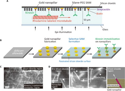
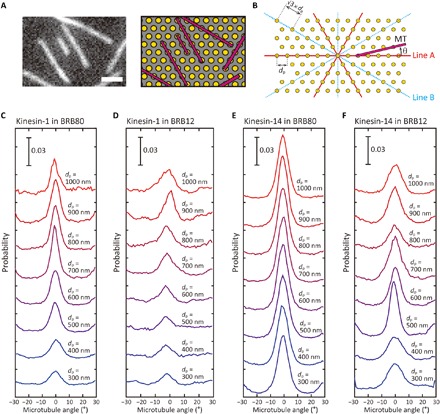
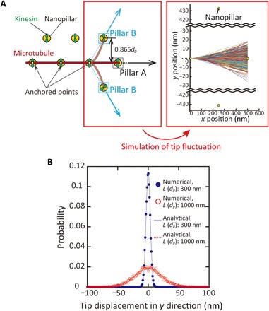
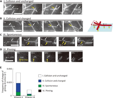
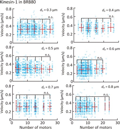
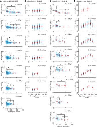
Kinesin is a motor protein that plays important roles in a variety of cellular functions. In vivo, multiple kinesin molecules are bound to cargo and work as a team to produce larger forces or higher speeds than a single kinesin. However, the coordination of kinesins remains poorly understood because of the experimental difficulty in controlling the number and arrangement of kinesins, which are considered to affect their coordination. Here, we report that both the number and spacing significantly influence the velocity of microtubules driven by nonprocessive kinesin-14 (Ncd), whereas neither the number nor the spacing changes the velocity in the case of highly processive kinesin-1. This result was realized by the optimum nanopatterning method of kinesins that enables immobilization of a single kinesin on a nanopillar. Our proposed method enables us to study the individual effects of the number and spacing of motors on the collective dynamics of multiple motors.


Figures

Fig. 1
Fig. 1. Nanopatterning of kinesin molecules by selective immobilization of kinesin on gold nanopillars.
(A) Schematic illustration of nanopatterned kinesin molecules. (B) The experimental procedure of nanopatterning of kinesin molecules. (C) Fluorescence image of microtubules gliding at the boundary of the passivated silicon dioxide region and the kinesin-1–patterned region on gold nanopillars. Scale bar, 10 μm. (D) Sequential images of a microtubule at the boundary between the nanopillar region and silicon dioxide region. Scale bar, 2 μm.
Fig. 2
Fig. 2. Alignment of microtubules on patterned kinesin molecules.
(A) Fluorescence image of aligned microtubules on patterned kinesin-1 (left) and corresponding illustration of microtubules and nanopillars (right). The spacing between the pillars, dp, is 500 nm. Scale bar, 2 μm. The illustration is not to scale. (B) The definition of the orientation angle of microtubules. dp indicates the designed spacing between pillars. (C to F) The distribution of angles between microtubules and line A on patterned kinesin-1 in (C) BRB80 and (D) BRB12 and on patterned kinesin-14 in (E) BRB80 and (F) BRB12.
Fig. 3
Fig. 3. Simulation of a fluctuating leading tip of microtubules.
(A) Schematic illustration of a fluctuating tip of the microtubule (left) and representative traces of the simulated shape of the tip (right). Traces were obtained by 500 runs of a simulation. (B) Probability density function of the displacement of the tip in y direction. L, the length of a free tip; dp, the spacing between pillars. n > 10,000.
Fig. 4
Fig. 4. Behavior of microtubules that changed travel direction on patterned kinesins.
(A to D) Representative sequential images of the microtubules. Scale bars, 5 μm (A) “I. Collision and unchanged”: A microtubule did not change direction after collision; (B) “II. Collision and changed”: A microtubule changed direction after a collision. The white arrows indicate the leading tip of the microtubule. The white dashed arrows show the direction of microtubule gliding. The yellow arrows indicate the point of collision; (C) “III. Spontaneous”: A microtubule spontaneously changed its gliding direction. The yellow arrows indicate the position in which the leading tip changed its direction; (D) “IV. Pinning”: A microtubule changed direction because the leading end was pinned. (E) Frequency of the events (I) to (IV) on patterned kinesin-1 and kinesin-14.
Fig. 5
Fig. 5. Effect of the number and spacing of kinesin-1 proteins on microtubule velocity in BRB80 buffer.
Blue open circles represent individual measurements, and red points represent the average velocity binned into five-motor intervals. Means ± SD; n.s., not significant (Steel-Dwass test).
Fig. 6
Fig. 6. Effect of the number and spacing of kinesin-14 proteins on microtubule velocity.
(A and B) Dependence of microtubule velocity on (A) the number of kinesin-14 proteins and (B) the spacing of kinesin-14 proteins in the BRB80 buffer. (A and B) Dependence of the microtubule velocity on (A) the number of kinesin-14 proteins and (B) the spacing of kinesin-14 proteins in the BRB12 buffer. Blue open circles represent individual measurements, and red circles represent the average velocity binned into five-motor intervals. Means ± SD; *P < 0.05 (Steel-Dwass test).

References

    1. Hirokawa N., Noda Y., Tanaka Y., Niwa S., Kinesin superfamily motor proteins and intracellular transport. Nat. Rev. Mol. Cell Biol. 10, 682–696 (2009). - PubMed
    1. Brust-Mascher I., Scholey J. M., Mitotic motors and chromosome segregation: The mechanism of anaphase B. Biochem. Soc. Trans. 39, 1149–1153 (2011). - PubMed
    1. Caruel M., Truskinovsky L., Physics of muscle contraction. Rep. Prog. Phys. 81, 036602 (2018). - PubMed
    1. Riedel-Kruse I. H., Hilfinger A., Howard J., Jülicher F., How molecular motors shape the flagellar beat. HFSP J. 1, 192–208 (2007). - PMC - PubMed
    1. Holzbaur E. L. F., Goldman Y. E., Coordination of molecular motors: From in vitro assays to intracellular dynamics. Curr. Opin. Cell Biol. 22, 4–13 (2010). - PMC - PubMed

Publication types