

Spinal epidural involvement in adult Langerhans cell histiocytosis (LCH): A case report
- PMID: 32011480
- PMCID: PMC7220126
- DOI: 10.1097/MD.0000000000018794
Spinal epidural involvement in adult Langerhans cell histiocytosis (LCH): A case report
Abstract
Rationale: Spinal involvement in adult Langerhans cell histiocytosis (LCH) is rare, and epidural involvement is unusual. LCH is mostly indistinguishable from other spinal lesions such as infection, lymphoma, and metastasis. So, it could be easily misdiagnosed without suspicion.
Patient concerns: We report a case of a 33-year-old man who complained of gait disturbance with weakness in both legs and severe back pain.
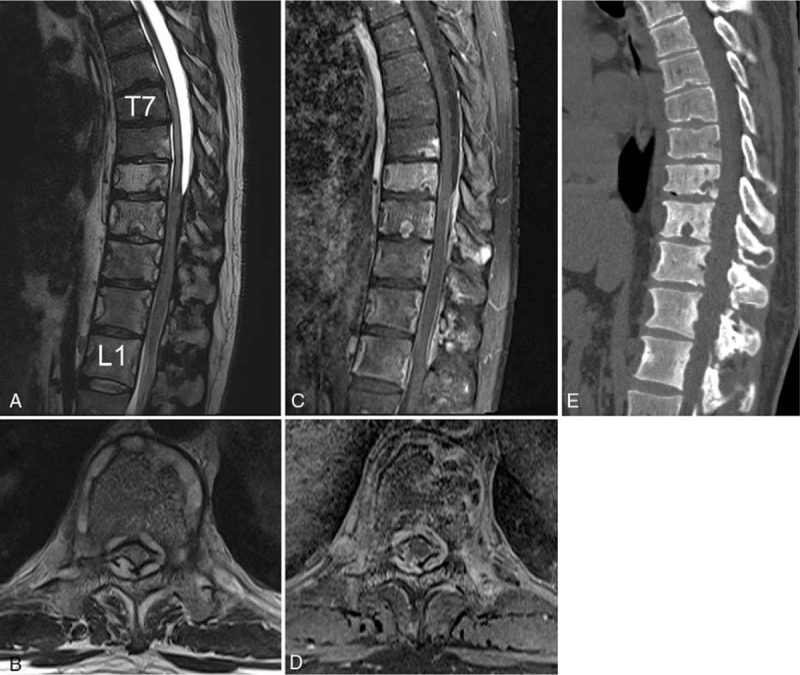
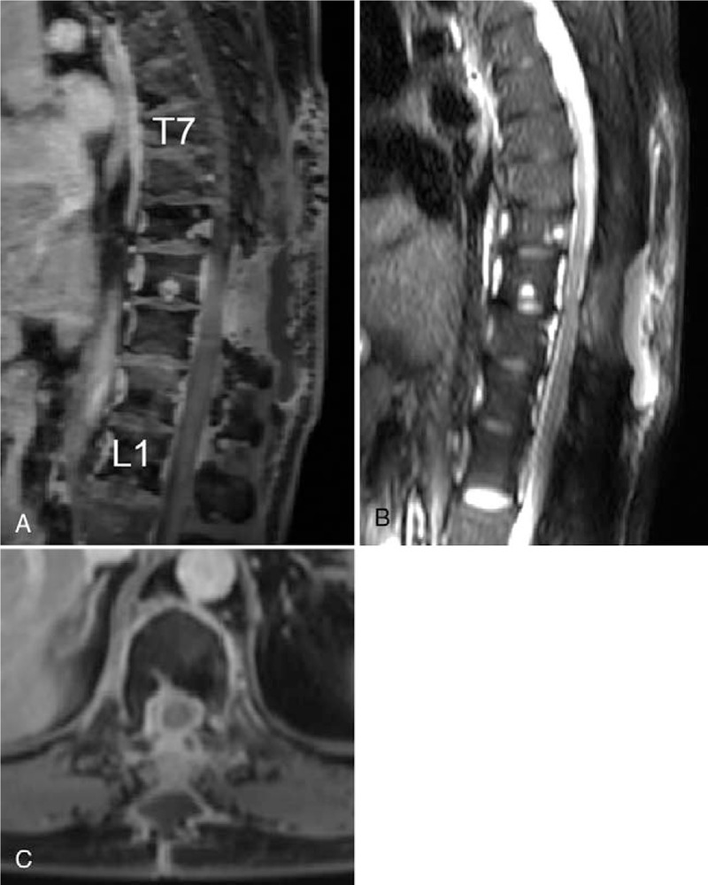
Diagnoses: A continuous enhancing epidural lesion with cord compression from the T7 to L1 level was detected in magnetic resonance imaging. Laboratory analysis indicated the possibility of spinal infectious disease. We assumed that the lesion could be tuberculous spondylitis.
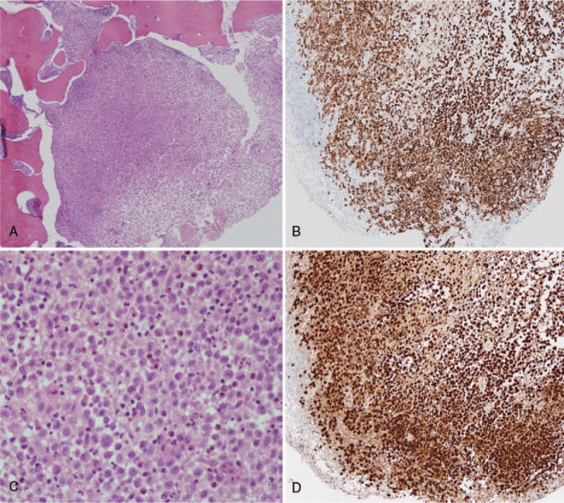
Interventions and outcomes: The patient underwent posterior laminectomy with marginal excision of the epidural mass to relieve cord compression. Pathological examination confirmed the diagnosis of LCH. The 12-month follow-up evaluation revealed that the patient was neurologically intact and had no gait disturbance.
Lessons: This case report presents a patient with epidural LCH of the thoracic spinal cord, which can mimic spinal infections such as tuberculous spondylitis with abscess formation. Therefore, LCH could be considered as a possible diagnosis when a patient presents with features of infectious spondylitis with vertebral involvement.
Conflict of interest statement
The authors report no conflicts of interest.
Figures
References
-
- Monsereenusorn C, Rodriguez-Galindo C. Clinical characteristics and treatment of langerhans cell histiocytosis. Hematol Oncol Clin North Am 2015;29:853–73. - PubMed
-
- Jiang L, Liu ZJ, Liu XG, et al. Langerhans cell histiocytosis of the cervical spine: a single Chinese institution experience with thirty cases. Spine (Phila Pa 1976) 2010;35:E8–15. - PubMed
-
- Yeom JS, Lee CK, Shin HY, et al. Langerhans’ cell histiocytosis of the spine. Analysis of twenty-three cases. Spine (Phila Pa 1976) 1999;24:1740–9. - PubMed
-
- Huang WD, Yang XH, Wu ZP, et al. Langerhans cell histiocytosis of spine: a comparative study of clinical, imaging features, and diagnosis in children, adolescents, and adults. Spine J 2013;13:1108–17. - PubMed
Publication types
MeSH terms
LinkOut - more resources
Full Text Sources
