. 2020 Feb 28;477(4):773-786.
doi: 10.1042/BCJ20190733.

Full-length NF-κB repressing factor contains an XRN2 binding domain

Affiliations

Full-length NF-κB repressing factor contains an XRN2 binding domain

Jana Alexandrova et al. Biochem J. .

Abstract

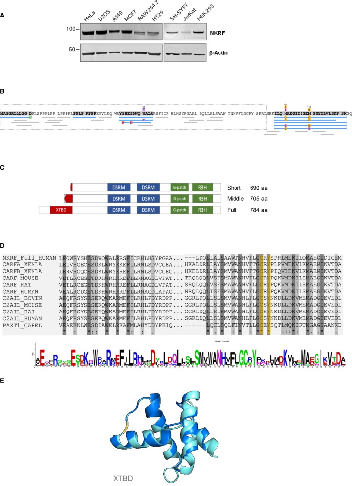
NF-κB repressing factor (NKRF) was recently identified as an RNA binding protein that together with its associated proteins, the 5'-3' exonuclease XRN2 and the helicase DHX15, is required to process the precursor ribosomal RNA. XRN2 is a multi-functional ribonuclease that is also involved in processing mRNAs, tRNAs and lncRNAs. The activity and stability of XRN2 are controlled by its binding partners, PAXT-1, CDKN2AIP and CDKN2AIPNL. In each case, these proteins interact with XRN2 via an XRN2 binding domain (XTBD), the structure and mode of action of which is highly conserved. Rather surprisingly, although NKRF interacts directly with XRN2, it was not predicted to contain such a domain, and NKRF's interaction with XRN2 was therefore unexplained. We have identified an alternative upstream AUG start codon within the transcript that encodes NKRF and demonstrate that the full-length form of NKRF contains an XTBD that is conserved across species. Our data suggest that NKRF is tethered in the nucleolus by binding directly to rRNA and that the XTBD in the N-terminal extension of NKRF is essential for the retention of XRN2 in this sub-organelle. Thus, we propose NKRF regulates the early steps of pre-rRNA processing during ribosome biogenesis by controlling the spatial distribution of XRN2 and our data provide further support for the XTBD as an XRN2 interacting motif.

Keywords: RNA-binding proteins; XRN2; XTBD; protein synthesis; ribosome biogenesis.


Conflict of interest statement

The authors declare that there are no competing interests associated with the manuscript.

Figures

Figure 1.
Figure 1.. The full-length NKRF transcript encodes a protein of 784 amino acids.
(A) Schematic representation of the NKRF mRNA variants identified in the sequence databases. The three main exons are represented in orange, blue and grey. AUG0 is initiation codon for the short NKRF protein; AUG1 is initiation codon for the version derived from mRNA variant 1 that contains an additional 15 amino acids; AUG2 is the initiation codon for the full-length NKRF identified herein. (B) Western analysis of U-2 OS cell extracts reveals that NKRF protein migrates at 88 kDa and not at the predicted molecular mass of 77 kDa. RNAi-mediated depletion was used to reduce the cellular abundance of NKRF using two NKRF siRNAs (A,B) and a control siRNA (ctrl). Western analysis confirmed that the 88 kDa protein is depleted by both NKRF siRNAs demonstrating that the antibody specifically recognises NKRF protein. (C) The sequences of NKRF from different mammals were compared. Green denotes the upstream initiation codon AUG2, and blue the cytosine nucleotide missing from the human reference genomic sequence. (D) Ribosome profiling experiments were analysed and visualised using the Genome Wide Information on Protein Synthesis tool (GWIPS-viz) [31] to determine the position of translating ribosomes on the NKRF mRNA. The global aggregate of ribosome footprint reads from 43 ribosome profiling studies was plotted against the NKRF gene sequence to generate a ribosome density map of elongating ribosomes on the NKRF gene (red profile). As expected, ribosome density can be detected in the previously annotated coding exons 2 and 3. However, ribosome density also extends into and upstream of the previously described exon 1, which was assigned as non-coding, as far as the new initiation codon AUG2 (red profile). Enrichment of ribosome density can be seen at AUG2, but not at AUG1 or AUG0, after treatment of cells with homoharrintonine (blue plot) [18]. RNA-seq reads (green profile) indicate that the NKRF mRNA extends further upstream than the RefSeq mRNA database entry (NM_0017544.3). The grey dotted lines highlight the position of AUG2, AUG1 and AUG0.
Figure 2.
Figure 2.. Full-length NKRF contains an XRN2 binding domain (XTBD).
(A) Cell extracts from HeLa, U-2 OS, A549, MCF7, RAW264.7, HT29, SH-SY5Y, Jurkat and HEK293 cells were separated by SDS PAGE and subjected to western analysis using an antibody against NKRF. Actin was used as a loading control. (B) NKRF was immunoprecipitated and subjected to mass spectrometry which identified full-length NKRF (NKRF_full) containing an intact XTBD domain. The sequence of the N-terminus of the extended NKRF is depicted with the sequence that is unique to NKRF_full in the grey dotted box. The three sequences that are unique to NKRF_full that were identified by mass spectrometry are highlighted in grey. Peptides that were identified in these experiments are represented as blue lines. (C) A schematic presentation of NKRF proteins is shown representing (i) the short isoform of 690 amino acids, (ii) the middle isoform of 705 amino acids, and (iii) the full-length protein of 784 amino acids. All isoforms of NKRF contain three RNA binding domains: G-Patch, RH3, and double strand RNA binding domain. Only full-length NKRF contains the intact XRN2 binding domain (red). (D) The NKRF XRN2 binding domain (XTBD) contains functionally important conserved residues. Sequence comparison of XTBD from CDKN2AIP (from mouse, rat and human), C2AIL (CDKN2AIPNL) (from mouse, rat, bovine and human), PAXT-1 (C. elegans) and human full-length NKRF shows a high degree of conservation and particularly residues Cys54 and Tyr56 (orange), which are essential for the interaction with XRN2. (E) A structural model of the NKRF XTBD domain was generated using RaptorX [32] and this structure was superimposed on the crystal structure of PAXT-1 using PyMol. The residues Cys54 and Tyr56, which are essential for the interaction with XRN2, are shown in orange.
Figure 3.
Figure 3.. The interaction between NKRF and XRN2 is independent of rRNA.
(A) U-2 OS cells were treated with the specific RNAPI inhibitor CX-5461 (CX) and cell extracts were generated from untreated and treated cells. Untreated cell lysates were incubated with either RNase or DNase for 2 h at 4°C. NKRF was then immunoprecipitated using an anti-NKRF antibody or the control IgG. The corresponding samples were separated by SDS–PAGE and immunoblotted with antibodies directed against NKRF and XRN2. Nucleolin was used as negative control. (B) U-2 OS cells were transfected with control siRNA (i) or siRNA targeting NKRF (ii) and after 72 h cells were fixed and permeabilised. XRN2 cellular distribution was determined by immunolocalisation. XRN2 is visualised in green; Hoechst (blue) and fibrillarin (red) were used as nuclear and nucleolar markers, respectively.
Figure 4.
Figure 4.. Co-localization of NKRF and XRN2 is dependent on RNAPI activity.
(A) RNA interactome capture was performed on U-2 OS cells that were either untreated or treated with a low dose of actinomycin or 2.5 µM CX-5641 to inhibit RNAPI. The samples were immunoblotted and probed with antibodies against XRN2 and NKRF. Polypyrimidine tract binding protein (PTB) was used as a control. (B) U-2 OS cells were either untreated or exposed to 2.5 µM CX-5461 and immunolocalization was performed. The subcellular distribution of either NKRF (green) or fibrillarin (red) was determined using specific antibodies. Hoechst (blue) and fibrillarin (red) were used as nuclear and nucleolar marker, respectively. (C) U-2 OS cells either untreated or exposed to 2.5 µM CX-5461 and immunolocalization was performed. The subcellular distribution of either XRN2 (green) or fibrillarin (red) was determined using specific antibodies. Hoechst (blue) and fibrillarin (red) were used as nuclear and nucleolar marker, respectively.
Figure 5.
Figure 5.. Full-length NKRF containing the XTBD interacts with XRN2.
(A) Schematic to show the three variants of NKRF that were expressed in U-2 OS cells. The expressed proteins are tagged at the N-terminus with a 3xFLAG tag. (B) U-2 OS cells were transiently transfected with the constructs expressing short, middle or full-length NKRF containing an N-terminal expressed FLAG tag. Cell extracts were generated and proteins were immunoprecipitated using anti-FLAG antibody. IgG was used as negative control. Isolated proteins were separated by SDS–PAGE and subjected to western analysis.
Figure 6.
Figure 6.. Full-length NKRF containing the XTBD restores the nucleolar localization of XRN2.
(A) NKRF short, middle and full-length U-2 OS stable cell lines were transfected with an siRNA to the NKRF open reading frame (siORF), which would target both the endogenous and expression versions of the proteins, or the NKRF 3′UTR (si3′UTR), which would only target endogenous NKRF. After 72 h, the expression of the NKRF isoforms was induced with tetracycline for a further 24 h. Cell extracts were generated and immunoblotted with antibodies against either FLAG or NKRF. Endogenous NKRF is marked (red asterisk). (B) NKRF short (i), middle (ii) and full-length (iii) U-2 OS stable cell lines were transfected with siRNAs targeting the 3′UTR of endogenous NKRF. After 72 h, expression of the FLAG-tagged versions of NKRF was induced for 24 h with tetracycline. Cells were fixed and permeabilised and the subcellular distribution of XRN2 (red) was determined using immunolocalization. NKRF (green) was visualised using anti-FLAG antibodies. Nuclei were stained with Hoechst (blue). (C) The data in (B) were quantified by counting ∼100 cells per condition to assess the XRN2 distribution in the nucleoli after expression of the different version of NKRF. The percentage of cells where XRN2 was present in the nucleoli was calculated (n = 3, error bars represent SD, and the significance was calculated using Fisher's exact test).

References

    1. de la Cruz J., Karbstein K. and Woolford J.L. Jr (2015) Functions of ribosomal proteins in assembly of eukaryotic ribosomes in vivo. Annu. Rev. Biochem. 84, 93–129 10.1146/annurev-biochem-060614-033917 - DOI - PMC - PubMed
    1. Bohnsack K.E. and Bohnsack M.T. (2019) Uncovering the assembly pathway of human ribosomes and its emerging links to disease. EMBO J. 38, e100278 10.15252/embj.2018100278 - DOI - PMC - PubMed
    1. Monaco P.L., Marcel V., Diaz J.J. and Catez F. (2018) 2′-O-methylation of ribosomal RNA: towards an epitranscriptomic control of translation? Biomolecules 8, E106 10.3390/biom8040106 - DOI - PMC - PubMed
    1. Tafforeau L., Zorbas C., Langhendries J.L., Mullineux S.T., Stamatopoulou V., Mullier R. et al. (2013) The complexity of human ribosome biogenesis revealed by systematic nucleolar screening of Pre-rRNA processing factors. Mol. Cell 51, 539–551 10.1016/j.molcel.2013.08.011 - DOI - PubMed
    1. Turowski T.W. and Tollervey D. (2015) Cotranscriptional events in eukaryotic ribosome synthesis. Wiley Interdiscip. Rev. RNA 6, 129–139 10.1002/wrna.1263 - DOI - PubMed

Publication types

LinkOut - more resources