. 2020 Feb 3;14(2):e0007948.
doi: 10.1371/journal.pntd.0007948. eCollection 2020 Feb.

Aedes cadherin receptor that mediates Bacillus thuringiensis Cry11A toxicity is essential for mosquito development

Affiliations

Aedes cadherin receptor that mediates Bacillus thuringiensis Cry11A toxicity is essential for mosquito development

Jianwu Chen et al. PLoS Negl Trop Dis. .

Abstract

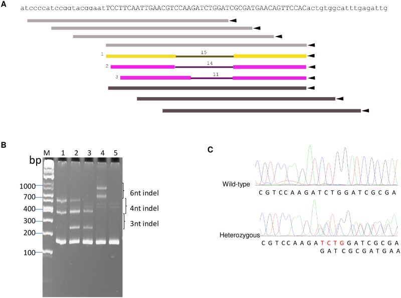
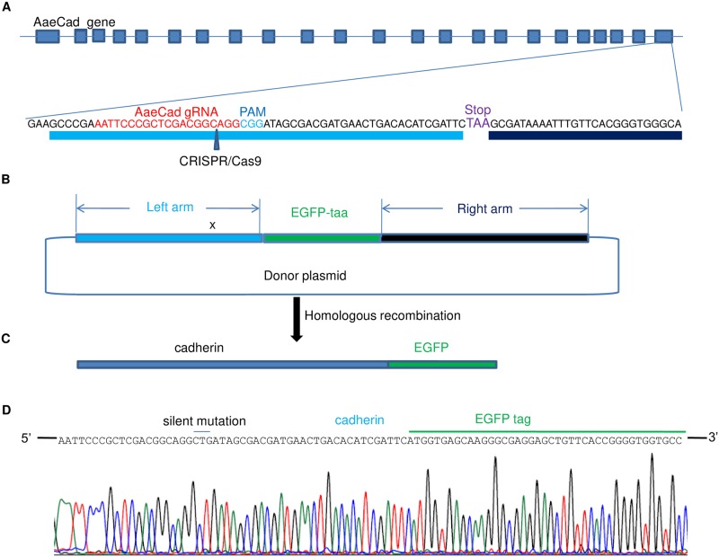
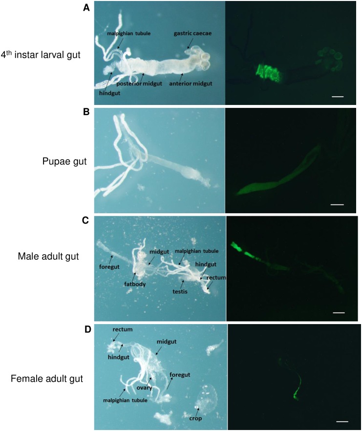
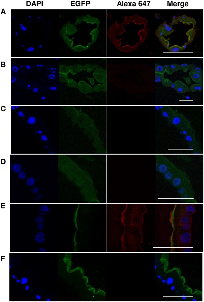
Aedes cadherin (AaeCad, AAEL024535) has been characterized as a receptor for Bacillus thuringiensis subsp. israelensis (Bti) Cry11A toxins. However, its role in development is still unknown. In this study, we modified the cadherin gene using ZFN and TALEN. Even though we obtained heterozygous deletions, no homozygous mutants were viable. Because ZFN and TALEN have lower off-targets than CRISPR/Cas9, we conclude the cadherin gene is essential for Aedes development. In contrast, in lepidopteran insects loss of a homologous cadherin does not appear to be lethal, since homozygous mutants are viable. To analyze the role of AaeCad in vivo, we tagged this protein with EGFP using CRISPR-Cas9-mediated homologous recombination and obtained a homozygous AaeCad-EGFP line. Addition of Aedes Rad51 mRNA enhanced the rate of recombination. We then examined AaeCad protein expression in most tissues and protein dynamics during mosquito development. We observe that AaeCad is expressed in larval and adult midgut-specific manner and its expression pattern changed during the mosquito development. Confocal images showed AaeCad has high expression in larval caecae and posterior midgut, and also in adult midgut. Expression of AaeCad is observed primarily in the apical membranes of epithelial cells, and not in cell-cell junctions. The expression pattern observed suggests AaeCad does not appear to play a role in these junctions. However, we cannot exclude its role beyond cell-cell adhesion in the midgut. We also observed that Cry11A bound to the apical side of larval gastric caecae and posterior midgut cells exactly where AaeCad-EGFP was expressed. Their co-localization suggests that AaeCad is indeed a receptor for the Cry11A toxin. Using this mosquito line we also observed that low doses of Cry11A toxin caused the cells to slough off membranes, which likely represents a defense mechanism, to limit cell damage from Cry11A toxin pores formed in the cell membrane.


Conflict of interest statement

The authors have declared that no competing interests exist.

Figures

Fig 1
Fig 1. Use of ZFN results in efficient editing of the AaeCad gene.
A. A ZFN construct with zinc finger nuclease binding site (in black, uppercase) and target site (in red, lower case). B. After G1 mosquitoes were genotyped and the generated data was analyzed and plotted. Profiling of the deletion length and the mutation rate is shown for a representative group of G1 mosquitoes. C. A chromatogram of a ZFN-targeted AaeCad fragment from wild-type Orlando strain mosquitoes and heterozygous mutant with 4nt-deletion. For ZFN-targeted AaeCad fragment from the heterozygous mutant, the overlapping chromatography peaks are displayed after the 4nt deletion.
Fig 2
Fig 2. TALEN mediated knockout of the AaeCad gene.
A. In this TALEN design schematics shows AaeCad gene sequence targeted by TALEN. B. G2 mosquitoes were genotyped by native heteroduplex mobility assay (HMA). Four out of 117 mosquito groups analyzed had 3nt, 4nt, 6nt or 3nt & 4nt deletions, respectively. Lane M, DNA marker; Lane 1, sample mosquito #31M with 4nt indel; Lane 2, sample #32M with 3nt and 4nt indel; Lane 3, sample #41 with 3nt indel; Lane 4, sample #70F with 6nt indel; Lane 5, sample #24F with no indel. C. PCR product sequencing from a heterozygous mutant displayed overlapping chromatogram peaks, but those from wild-type Orlando strain mosquito showed single chromatogram peaks.
Fig 3
Fig 3. CRISPR-Cas9 mediated EGFP tagging at the 3’ end of the last coding exon of AaeCad gene results in an in frame fusion.
A. sgRNA (in red) and PAM sequences (in blue) were designed before the stop codon (in purple) on the last exon of AaeCad gene. B. This donor plasmid for homologous recombination (HR) contains 1000bp of left arm, 720 bp of EGFP ORF and 1000bp of right arm. To prevent Cas9 cleavage on the left arm in the plasmid, a silent mutation (CGG → CTG) was introduced into the PAM sequence on the left arm. C. When the homologous recombination occurred, the EGFP was introduced immediately after the AaeCad gene with the aid of the left and right arms. D. The sequencing results of gDNA showed the AaeCad gene had been successfully tagged with EGFP and no mutation was introduced into the AaeCad gene except for the silent mutation that was intentionally introduced.
Fig 4
Fig 4. Visualization of AaeCad-EGFP in whole mounts of dissected guts from larva, pupa and adult male and female homozygous Aedes.
A. AaeCad protein localization in early 4th instar larval gut. Expression is observed primarily in the gastric caecae and the posterior midgut. B. AaeCad protein expression in pupal gut. C. AaeCad protein localization in adult male gut; D. AaeCad protein localization in adult female gut. Expression in both the male and female was observed primarily in the foregut. Since the tissues were in PBS buffer the samples move ever so slightly under the different filters, preventing use of the merge function. Bar: 50μm.
Fig 5
Fig 5. AaeCad protein is localized primarily in gastric caecae and posterior midgut of early 4th instar larvae.
A. Whole mount images under low magnification showed that strong AaeCad-EGFP expression in the gastric caecae (GC) and posterior midgut cell membrane. B. Cross section images showed high AaeCad-EGFP expression in the gastric caecae (GC) and posterior midgut and low expression in the anterior midgut. In the gastric caecae and midgut, EGFP-tagged AaeCad is specifically expressed in the epithelial cell membrane. DAPI stains the nucleus. All images were collected using an SP5 Inverted confocal microscope. Bar: 50μm.
Fig 6
Fig 6. An anti-AaeCad polyclonal detects the EGFP-tagged AaeCad.
Anti-AaeCad polyclonal antibody could detect the EGFP-tagged AaeCad in gastric caecae (A) and posterior midgut (E), but not in the anterior midgut (C). Whereas, when only the Alexa 647-labeled antibody was used, no fluorescence was detected in the gastric caecae (B), anterior midgut (D) and posterior midgut (F). Bar: 50μm, but is 25μm in panel E.
Fig 7
Fig 7. EGFP-tagged AaeCad co-localizes in the larval gut cells with the Cry11Aa toxin.
The Cry11A toxin bound to the AaeCad-EGFP in the gastric caecae (A) and posterior midgut (E), but not in the anterior midgut (C). However, when the Cry11A toxin was absent, the fluorescence could not be observed in the gastric caecae (B), anterior midgut (D) and posterior midgut (F). Bar: 50μm, but is 25μm in panels E and F.
Fig 8
Fig 8. Larval exposure to low level Cry11A toxin concentrations disrupts mosquito midgut cells and causes these cells to shed its cell membrane.
The larval midgut cell morphology was observed under a confocal microscope after the mosquitos were treated at the LC10 dose for 1 hr (A), 4 hr (B), 8 hr (C) and 16 hr (D). Changes in cell conformation were observed with Armadillo staining in cadherin-EGFP mosquitoes (E, F and G) at 18h. The same image is observed with DAPI, EFGP (cadherin) and Alexa 555 (Amardillo). Significant AaeCad-EGFP and Amardillo signaling is also observed in the intracellular compartments. Bar: 50μm.

Similar articles

Cited by

References

    1. Kyle JL, Harris E (2008) Global spread and persistence of dengue. Annu Rev Microbiol 62: 71–92. 10.1146/annurev.micro.62.081307.163005 - DOI - PubMed
    1. Zhang Q, Hua G, Adang MJ (2017) Effects and mechanisms of Bacillus thuringiensis crystal toxins for mosquito larvae. Insect Sci 24: 714–729. 10.1111/1744-7917.12401 - DOI - PubMed
    1. Ben-Dov E (2014) Bacillus thuringiensis subsp. israelensis and its dipteran-specific toxins. Toxins (Basel) 6: 1222–1243. - PMC - PubMed
    1. Resh VH, Leveque C, Statzner B (2004) Long-term, large-scale biomonitoring of the unknown: assessing the effects of insecticides to control river blindness (onchocerciasis) in West Africa. Annu Rev Entomol 49: 115–139. 10.1146/annurev.ento.49.061802.123231 - DOI - PubMed
    1. Berry C, O'Neil S, Ben-Dov E, Jones AF, Murphy L, et al. (2002) Complete sequence and organization of pBtoxis, the toxin-coding plasmid of Bacillus thuringiensis subsp. israelensis. Appl Environ Microbiol 68: 5082–5095. 10.1128/AEM.68.10.5082-5095.2002 - DOI - PMC - PubMed

Publication types

MeSH terms