. 2020 Feb 3;15(2):e0228685.
doi: 10.1371/journal.pone.0228685. eCollection 2020.

Roles of the vestibular system in obesity and impaired glucose metabolism in high-fat diet-fed mice

Affiliations

Roles of the vestibular system in obesity and impaired glucose metabolism in high-fat diet-fed mice

Naoyuki Kawao et al. PLoS One. .

Abstract

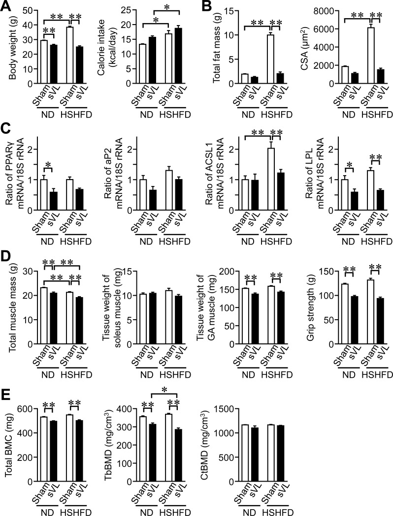
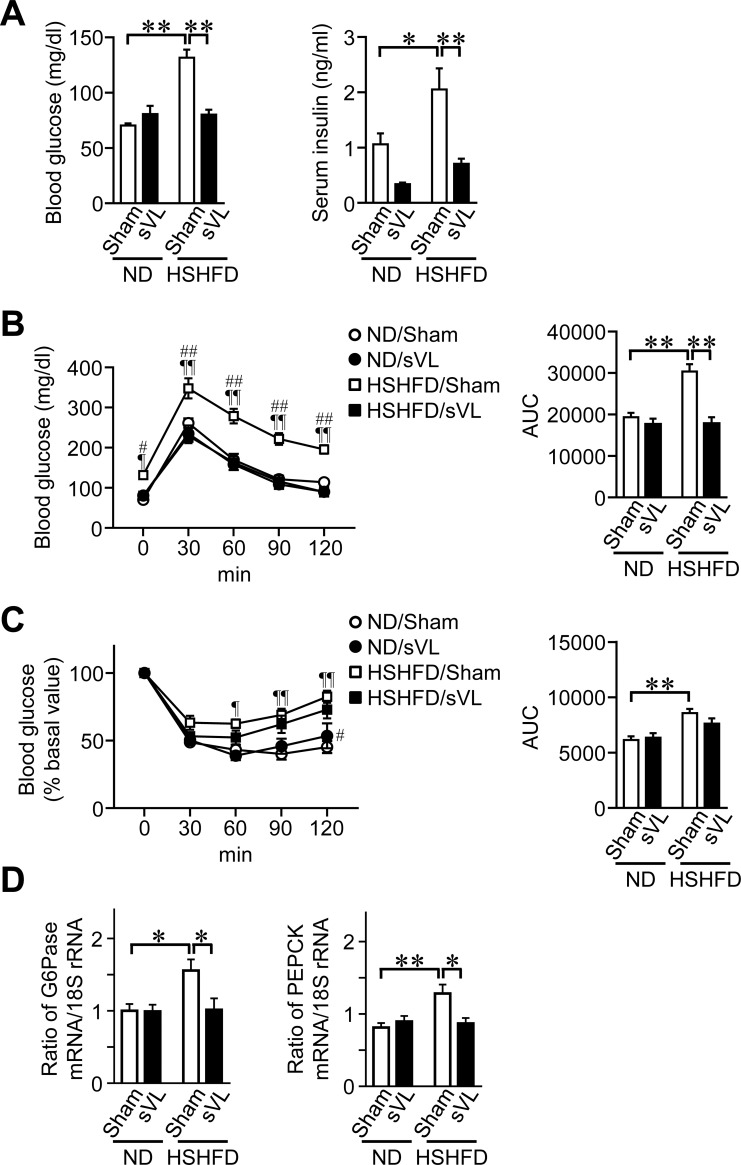
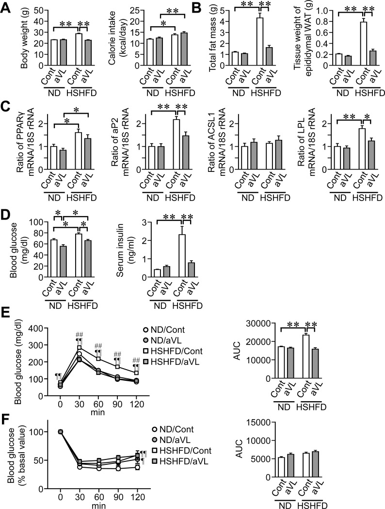
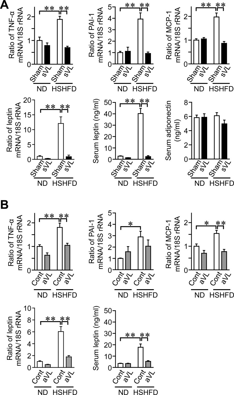
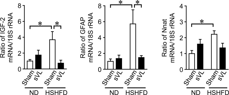
The vestibular system controls balance, posture, blood pressure, and gaze. However, the roles of the vestibular system in energy and glucose metabolism remain unknown. We herein examined the roles of the vestibular system in obesity and impaired glucose metabolism using mice with vestibular lesions (VL) fed a high-sucrose/high-fat diet (HSHFD). VL was induced by surgery or arsenic. VL significantly suppressed body fat enhanced by HSHFD in mice. Glucose intolerance was improved by VL in mice fed HSHFD. VL blunted the levels of adipogenic factors and pro-inflammatory adipokines elevated by HSHFD in the epididymal white adipose tissue of mice. A β-blocker antagonized body fat and glucose intolerance enhanced by HSHFD in mice. The results of an RNA sequencing analysis showed that HSHFD induced alterations in genes, such as insulin-like growth factor-2 and glial fibrillary acidic protein, in the vestibular nuclei of mice through the vestibular system. In conclusion, we herein demonstrated that the dysregulation of the vestibular system influences an obese state and impaired glucose metabolism induced by HSHFD in mice. The vestibular system may contribute to the regulation of set points under excess energy conditions.


Conflict of interest statement

The authors have declared that no competing interests exist.

Figures

Fig 1
Fig 1. Effects of sVL on body weight and composition in mice fed HSHFD for 8 weeks.
(A) Data on body weight and calorie intake from sham surgery and sVL mice fed ND or HSHFD. Body weight was measured 8 weeks after ND or HSHFD feeding. Food intake was collected for 3 days on days 54 to 56 after ND or HSHFD feeding was started and shown as a representative of the average daily calorie intake. (B) Fat mass in the whole body of sham surgery and sVL mice was assessed by QCT 8 weeks after ND or HSHFD feeding was started. The cross-sectional area (CSA) of adipocytes in the white epididymal adipose tissue (WAT) of sham surgery and sVL mice 8 weeks after ND or HSHFD feeding was started. (C) Total RNA was extracted from the epididymal WAT of sham surgery and sVL mice 8 weeks after ND or HSHFD feeding was started. A real-time PCR analysis was then performed. Data are expressed relative to the levels of 18S rRNA. (D) Muscle mass in the whole body of sham surgery and sVL mice was assessed by QCT 8 weeks after ND or HSHFD feeding was started. The tissue weights of the soleus and gastrocnemius (GA) muscles were measured 8 weeks after ND or HSHFD feeding. The grip strengths of the four limbs were measured by a grip strength meter in sham surgery and sVL mice 8 weeks after ND or HSHFD feeding was started. (E) Total BMC, trabecular (Tb) BMD, and cortical (Ct) BMD in the tibia of sham surgery and sVL mice were assessed by QCT 8 weeks after ND or HSHFD feeding was started. *P < 0.05 and **P < 0.01 (Tukey-Kramer test). Data represent the mean ± SEM of 8 mice in each group.
Fig 2
Fig 2. Effects of sVL on glucose metabolism in mice fed HSHFD for 8 weeks.
(A) Fasting blood glucose and serum insulin levels were measured 8 weeks after ND or HSHFD feeding was started. (B, C) Responses of blood glucose to a single intraperitoneal injection of glucose (B) and insulin (C) in sham surgery and sVL mice 8 weeks after ND or HSHFD feeding was started. The area under the curve (AUC) for 120 min was calculated. (D) Total RNA was extracted from liver tissues of sham surgery and sVL mice 8 weeks after ND or HSHFD feeding was started. A real-time PCR analysis was then performed. Data are expressed relative to 18S rRNA levels. *P < 0.05 and **P < 0.01; ¶P < 0.05 and ¶¶P < 0.01, vs ND/Sham; #P < 0.05 and ##P < 0.01, vs HSHFD/sVL (Tukey-Kramer test). Data represent the mean ± SEM of 8 mice in each group.
Fig 3
Fig 3. Effects of aVL on fat mass and glucose metabolism in mice fed HSHFD for 4 weeks.
(A) Data on body weight and calorie intake from control (Cont) and aVL mice fed ND or HSHFD. Body weight was measured 4 weeks after ND or HSHFD feeding. Food intake was collected for 3 days on days 26 to 28 after ND or HSHFD feeding was started and shown as a representative of the average daily calorie intake. (B) Fat mass in the whole body of control and aVL mice was assessed by QCT 4 weeks after ND or HSHFD feeding was started. The tissue weight of epididymal white adipose tissue (WAT) was measured 4 weeks after ND or HSHFD feeding. (C) Total RNA was extracted from the epididymal WAT of control and aVL mice 4 weeks after ND or HSHFD feeding was started. A real-time PCR analysis was then performed. Data are expressed relative to the levels of 18S rRNA. (D) Fasting blood glucose and insulin levels were measured 4 weeks after ND or HSHFD feeding was started. (E, F) Responses of blood glucose to a single intraperitoneal injection of glucose (E) and insulin (F) in control and aVL mice 4 weeks after ND or HSHFD feeding was started. The area under the curve (AUC) for 120 min was calculated. *P < 0.05 and **P < 0.01; ¶P < 0.05 and ¶¶P < 0.01, vs ND/Sham; ##P < 0.01, vs HSHFD/aVL (Tukey-Kramer test). Data represent the mean ± SEM of 8 mice in each group.
Fig 4
Fig 4. Effects of sVL and aVL on adipokine levels in mice fed HSHFD.
(A) Total RNA was extracted from the epididymal WAT of sham surgery and sVL mice 8 weeks after ND or HSHFD feeding was started. A real-time PCR analysis was then performed. Data are expressed relative to 18S rRNA levels. Serum samples were collected from sham surgery and sVL mice 8 weeks after ND and HSHFD feeding was started. The quantification of serum leptin and adiponectin levels was performed. (B) Total RNA was extracted from the epididymal WAT of control (Cont) and aVL mice 4 weeks after ND or HSHFD feeding was started. A real-time PCR analysis was then performed. Data are expressed relative to the levels of 18S rRNA. Serum samples were collected from control and aVL mice 8 weeks after ND and HSHFD feeding was started. The quantification of serum leptin levels was performed. *P < 0.05 and **P < 0.01 (Tukey-Kramer test). Data represent the mean ± SEM of 8 mice in each group.
Fig 5
Fig 5. Effects of the propranolol treatment on fat mass and glucose metabolism in mice fed HSHFD for 8 weeks.
(A) Data on body weight and calorie intake from mice with or without propranolol (Propra). Body weight was measured 8 weeks after ND or HSHFD feeding. Food intake was collected for 3 days on days 54 to 56 after ND or HSHFD feeding was started and shown as a representative of the average daily calorie intake. (B) Fat mass in the whole body of mice with or without propranolol was assessed by QCT 8 weeks after ND or HSHFD feeding was started. The tissue weight of epididymal WAT was measured 8 weeks after ND or HSHFD feeding. (C) The cross-sectional area (CSA) of adipocytes in the epididymal WAT of mice with or without propranolol 8 weeks after ND or HSHFD feeding was started. (D) Total RNA was extracted from the epididymal WAT of mice with or without propranolol 8 weeks after ND or HSHFD feeding was started. A real-time PCR analysis was then performed. Data are expressed relative to the levels of 18S rRNA. (E) Fasting blood glucose and serum insulin levels were measured 8 weeks after ND or HSHFD feeding was started. (F, G) Responses of blood glucose to a single intraperitoneal injection of glucose (F) and insulin (G) in mice with or without propranolol 8 weeks after ND or HSHFD feeding was started. The area under the curve (AUC) for 120 min was calculated. Cont; control. *P < 0.05 and **P < 0.01; ¶P < 0.05 and ¶¶P < 0.01, vs ND/Control; #P < 0.05 and ##P < 0.01, vs HSHFD/Propranolol (Tukey-Kramer test). Data represent the mean ± SEM of 8 mice in each group.
Fig 6
Fig 6. Effects of sVL and HSHFD feeding on gene expression in vestibular nuclei of mice.
Total RNA was extracted from the vestibular nuclei of sham surgery and sVL mice 8 weeks after ND or HSHFD feeding was started. A real-time PCR analysis was then performed. Data are expressed relative to the levels of 18S rRNA. *P < 0.05 (Tukey-Kramer test). Data represent the mean ± SEM of 6 mice in each group.

References

    1. Clarke AH, Grigull J, Mueller R, Scherer H. The three-dimensional vestibulo-ocular reflex during prolonged microgravity. Exp Brain Res. 2000;134: 322–334. 10.1007/s002210000476 - DOI - PubMed
    1. Yates BJ, Bolton PS, Macefield VG. Vestibulo-sympathetic responses. Compr Physiol. 2014;4: 851–887. 10.1002/cphy.c130041 - DOI - PMC - PubMed
    1. Morita H, Abe C, Tanaka K. Long-term exposure to microgravity impairs vestibulo-cardiovascular reflex. Sci Rep. 2016;6: 33405 10.1038/srep33405 - DOI - PMC - PubMed
    1. Levasseur R, Sabatier JP, Etard O, Denise P, Reber A. Labyrinthectomy decreases bone mineral density in the femoral metaphysis in rats. J Vestib Res. 2004;14: 361–365. - PubMed
    1. Vignaux G, Ndong JD, Perrien DS, Elefteriou F. Inner ear vestibular signals regulate bone remodeling via the sympathetic nervous system. J Bone Miner Res. 2015;30: 1103–1111. 10.1002/jbmr.2426 - DOI - PMC - PubMed

Publication types