. 2020 Feb 3;15(2):e0228727.
doi: 10.1371/journal.pone.0228727. eCollection 2020.

Cell-free hemoglobin increases inflammation, lung apoptosis, and microvascular permeability in murine polymicrobial sepsis

Affiliations

Cell-free hemoglobin increases inflammation, lung apoptosis, and microvascular permeability in murine polymicrobial sepsis

Jamie E Meegan et al. PLoS One. .

Abstract

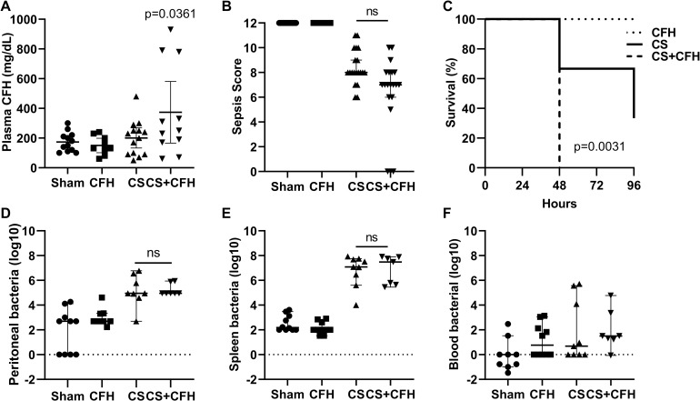
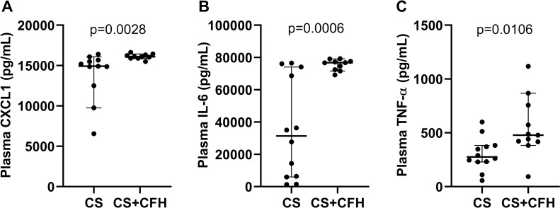
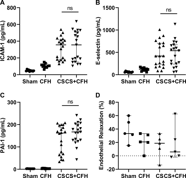
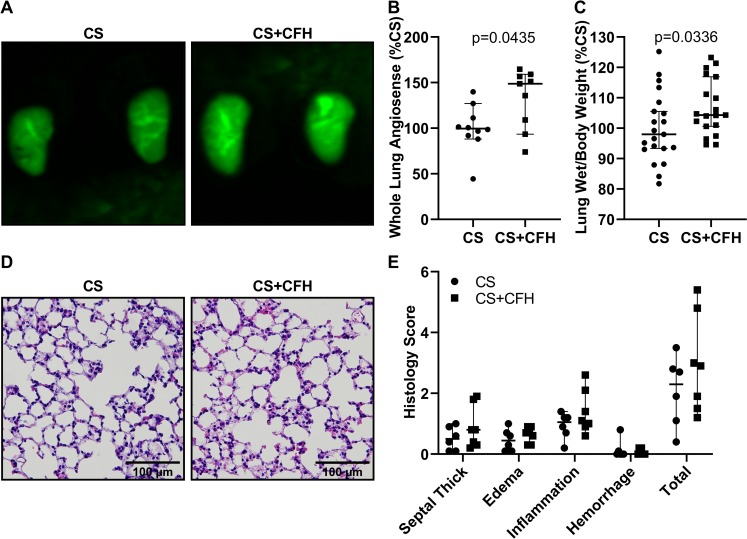
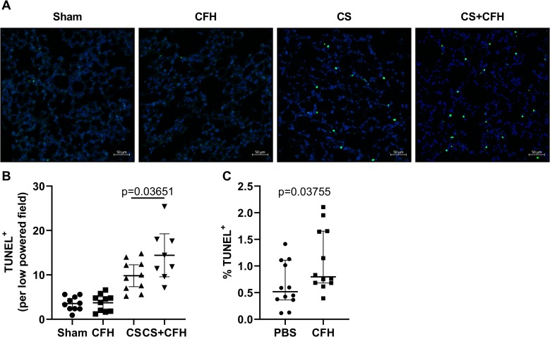
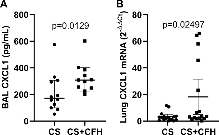
Increased endothelial permeability is central to the pathogenesis of sepsis and leads to organ dysfunction and death but the endogenous mechanisms that drive increased endothelial permeability are not completely understood. We previously reported that cell-free hemoglobin (CFH), elevated in 80% of patients with sepsis, increases lung microvascular permeability in an ex vivo human lung model and cultured endothelial cells. In this study, we augmented a murine model of polymicrobial sepsis with elevated circulating CFH to test the hypothesis that CFH increases microvascular endothelial permeability by inducing endothelial apoptosis. Mice were treated with an intraperitoneal injection of cecal slurry with or without a single intravenous injection of CFH. Severity of illness, mortality, systemic and lung inflammation, endothelial injury and dysfunction and lung apoptosis were measured at selected time points. We found that CFH added to CS increased sepsis mortality, plasma inflammatory cytokines as well as lung apoptosis, edema and inflammation without affecting large vessel reactivity or vascular injury marker concentrations. These results suggest that CFH is an endogenous mediator of increased endothelial permeability and apoptosis in sepsis and may be a promising therapeutic target.


Conflict of interest statement

The authors have declared that no competing interests exist.

Figures

Fig 1
Fig 1. Intravenous CFH augments mortality in sepsis.
To establish a clinically relevant model of sepsis with elevated circulating cell-free hemoglobin (CFH), we treated mice with an intravenous (IV) injection of CFH, an intraperitoneal (IP) injection of CS, simultaneous IV CFH and IP CS, or sham IV and IP injections. (A) After 24 hours, the CS+CFH group had the highest circulating CFH levels (n = 9–14, p = 0.0361 vs. Sham). The CS+CFH group had a (B) slightly lower sepsis score indicating more severe illness (n = 20–24, p = ns vs. CS) and (C) lower survival (n = 3–7, p = 0.0031). Bacterial counts measured in (D) peritoneal wash, (E) spleen homogenates and (F) blood were elevated in both CS and CS+CFH groups but did not differ between these two groups (n = 7–10).
Fig 2
Fig 2. Intravenous CFH augments systemic cytokine increases in murine sepsis.
Plasma cytokines were measured 24 hours after CS or CS+CFH treatment (n = 11–12). (A) CXCL1 (p = 0.0028), (B) IL-6 (p = 0.0006) and (C) TNF-α (p = 0.0106) were higher in CS+CFH versus CS alone.
Fig 3
Fig 3. CFH causes vascular injury in murine sepsis.
Markers of endothelial injury (n = 10–22), (A) ICAM-1, (B) E-selectin, and (C) PAI-1 were measured 24 hours after CS or CS+CFH treatment. All three markers are elevated in CS and CS+CFH groups but do not statistically differ from each other. (D) To test whether large vessel injury was induced by CFH, we measured aortic relaxation in response to acetylcholine in excised aortas 24 hours after treatment. CFH did not worsen aortic relaxation when combined with CS treatment (n = 5).
Fig 4
Fig 4. CFH increases lung edema formation in murine sepsis.
Lung microvascular permeability was measured by accumulation of fluorescent AngioSense in mouse lungs 24 hours after injection of sepsis. (A) Excised lungs were imaged using a high sensitivity camera. (B) Fluorescence intensity was quantified and was higher in CS+CFH versus CS alone (n = 9–10, p = 0.0435). (C) To measure lung fluid accumulation, we calculated lung wet weight to mouse body weight, expressed as a percent of CS treatment, and found that CS+CFH increases lung weight versus CS alone (n = 19–21, p = 0.0336). (D-E) Lung histology showed slightly increased septal thickening, edema, and inflammation in CS+CFH versus CS alone but was not statistically different (n = 6–7).
Fig 5
Fig 5. CFH increases lung endothelial apoptosis in sepsis.
(A) Formalin fixed mouse lung tissue was stained for TUNEL (green) with nuclear DAPI staining (blue). (B) The number of TUNEL positive cells was higher in CS+CFH versus CS alone by blinded quantification of low powered images (n = 8–10, p = 0.03651). (C) We also assessed apoptosis by TUNEL staining of HULECs in culture and found that CFH (1 mg/mL) treatment induced apoptosis at 4 hours (n = 12, p = 0.03755).
Fig 6
Fig 6. CFH increases lung inflammatory cytokine expression.
(A) CXCL1 measured by ELISA increased in mouse BAL collected 24 hours after induction of sepsis in CS+CFH versus CS alone (n = 11–12, p = 0.0129). (B) Lung CXCL1 expression measured by PCR in whole lungs at 24 hours also increased in CS+CFH versus CS alone (n = 16, p = 0.02497).
Fig 7
Fig 7. CFH increases lung oxidative injury.
(A) Whole lung F2-isoprostanes (n = 6, p = 0.0087) and (B) isofurans (n = 6, p = 0.0260) increased at 24 hours in CS+CFH versus CS alone.

References

    1. Lagu T, Rothberg MB, Shieh MS, Pekow PS, Steingrub JS, Lindenauer PK. Hospitalizations, costs, and outcomes of severe sepsis in the United States 2003 to 2007. Crit Care Med. 2012;40(3):754–61. 10.1097/CCM.0b013e318232db65 - DOI - PubMed
    1. Martin GS, Mannino DM, Eaton S, Moss M. The epidemiology of sepsis in the United States from 1979 through 2000. N Engl J Med. 2003;348(16):1546–54. 10.1056/NEJMoa022139 - DOI - PubMed
    1. Janz DR, Bastarache JA, Peterson JF, Sills G, Wickersham N, May AK, et al. Association between cell-free hemoglobin, acetaminophen, and mortality in patients with sepsis: an observational study. Crit Care Med. 2013;41(3):784–90. 10.1097/CCM.0b013e3182741a54 - DOI - PMC - PubMed
    1. Gladwin MT, Kanias T, Kim-Shapiro DB. Hemolysis and cell-free hemoglobin drive an intrinsic mechanism for human disease. J Clin Invest. 2012;122(4):1205–8. 10.1172/JCI62972 - DOI - PMC - PubMed
    1. Kuck JL, Bastarache JA, Shaver CM, Fessel JP, Dikalov SI, May JM, et al. Ascorbic acid attenuates endothelial permeability triggered by cell-free hemoglobin. Biochem Biophys Res Commun. 2017. - PMC - PubMed

Publication types