Review
. 2020 Jan 27;12(2):298.
doi: 10.3390/cancers12020298.

A Comprehensive Picture of Extracellular Vesicles and Their Contents. Molecular Transfer to Cancer Cells

Affiliations
Review

A Comprehensive Picture of Extracellular Vesicles and Their Contents. Molecular Transfer to Cancer Cells

Ancuta Jurj et al. Cancers (Basel). .

Abstract

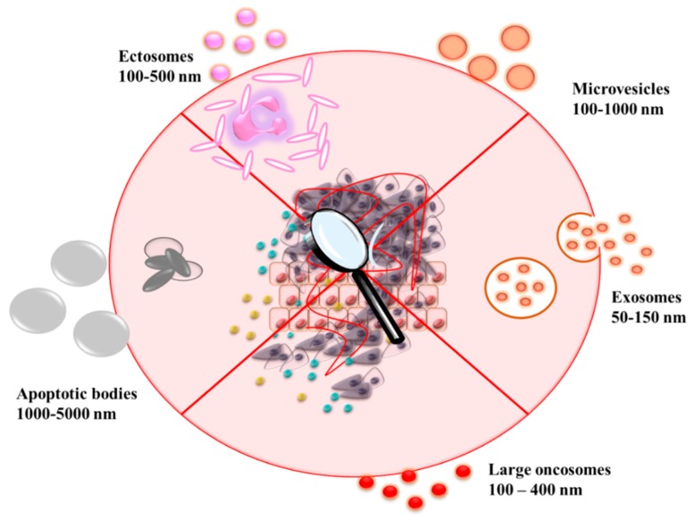
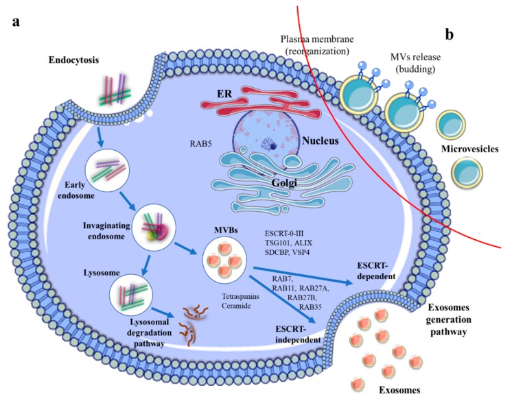
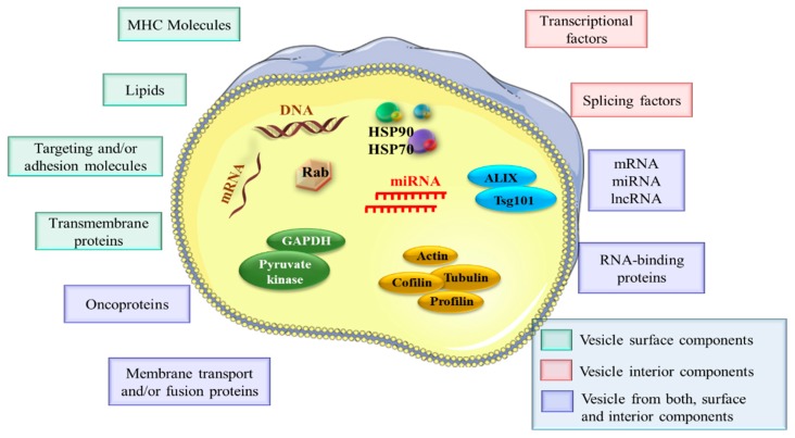
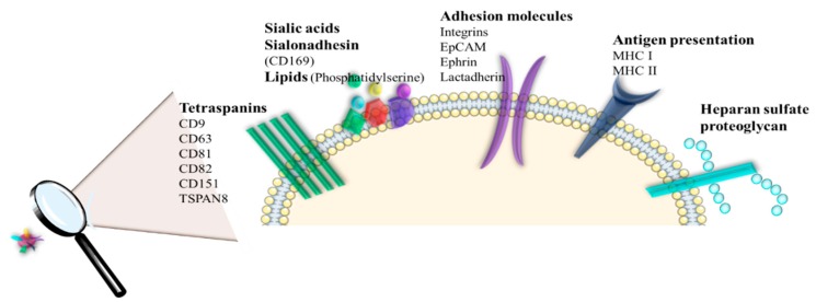
Critical processes such as growth, invasion, and metastasis of cancer cells are sustained via bidirectional cell-to-cell communication in tissue complex environments. Such communication involves the secretion of soluble factors by stromal cells and/or cancer cells within the tumor microenvironment (TME). Both stromal and cancer cells have been shown to export bilayer nanoparticles: encapsulated regulatory molecules that contribute to cell-to-cell communication. These nanoparticles are known as extracellular vesicles (EVs) being classified into exosomes, microvesicles, and apoptotic bodies. EVs carry a vast repertoire of molecules such as oncoproteins and oncopeptides, DNA fragments from parental to target cells, RNA species (mRNAs, microRNAs, and long non-coding RNA), and lipids, initiating phenotypic changes in TME. According to their specific cargo, EVs have crucial roles in several early and late processes associated with tumor development and metastasis. Emerging evidence suggests that EVs are being investigated for their implication in early cancer detection, monitoring cancer progression and chemotherapeutic response, and more relevant, the development of novel targeted therapeutics. In this study, we provide a comprehensive understanding of the biophysical properties and physiological functions of EVs, their implications in TME, and highlight the applicability of EVs for the development of cancer diagnostics and therapeutics.

Keywords: biogenesis; cancer; clinical implications; extracellular vesicles; function.


Conflict of interest statement

The authors declare no conflict of interest.

Figures

Figure 1
Figure 1
Various types of extracellular vesicles secreted from different cells, normal and tumor respectively.
Figure 2
Figure 2
Biogenesis mechanism of (a) exosomes and (b) MV, and their release processes which are coordinated through two different intracellular pathways, such as exosomes generation pathway and lysosomal degradation pathway. The exosomes formation starts with an active process, called endocytosis, where the cells internalized the material in the extracellular fluid to form internal vesicles (early and late endosomes). Through the inward budding of the late endosomal membrane, multivesicular bodies (MVBs) are formed. Moreover, MVBs can fuse with the lysosomes where their content is degraded or can traffic and fuse with the plasma membrane to release the content into the extracellular matrix. The exosomes generation pathway can be regulated through ESCRT-dependent or via ESCRT-independent pathway. Both processes, (MVBs fusion with the plasma membrane and exosomes release) use for regulation Rab GTPases (Rab7A < Rab11, Rab27A, Rab27B, and Rab35) and SNARE protein complex.
Figure 3
Figure 3
EV cargo profile. EVs deliver various bioactive molecules, including nucleic acids (DNA, mRNA, miRNAs (microRNAs), lncRNAs (long non-coding RNAs), specific proteins (oncoproteins), lipids, transcriptional factors, and RNA-binding proteins.
Figure 4
Figure 4
The presence of specific bioactive molecules on EV surface, which mediate the interactions between various ligands and receptors presented on the targeted cell surface (tetraspanins, adhesion molecules (integrins), lipids (phosphatidylserine), signaling receptors, molecules involved in antigen presentation and membrane trafficking (EpCAM—epithelial cell adhesion molecules, MHC—major histocompatibility complex).
Figure 5
Figure 5
EV internalization by recipient cells through different mechanisms, including direct membrane fusion, macropinocytosis, endocytosis (clathrin- and caveolae-dependent mechanism, lipid-raft-dependent endocytosis) and phagocytosis (PI3K-dependent, dynamic-dependent, and actin-polymerization-dependent mechanism). The presence of ligand-receptors present on recipient cell surface can elicit biological responses and can targeted EVs (MHC—major histocompatibility complex, TCR—T cell receptor, TRAIL—TNF-related apoptosis-inducing ligand, FASL—FAS antigen ligand, PD-L1—programmed cell death 1 ligand 1, FAS—apoptosis-mediating surface antigen, DR4—death receptor 4, DR5—death receptor 5, TNFR—TNF receptor, sTRAIL—soluble TRAIL, sFASL—soluble FASL, C3—complement component C3, C4—complement component C4, C5—complement component C5, PSGL1—P-selection glycoprotein ligand 1, ER—endoplasmic reticulum).
Figure 6
Figure 6
An overview of EV functions in TME cooperation. EVs derived from tumor cells act in an autocrine and paracrine manner. The interaction between tumor cells and other cells of TME through EVs may result in proliferation, tumor growth, metastasis, and drug resistance. EVs derived from tumor cells are involved in macrophage polarization, immune suppression, the transformation of fibroblast to cancer-associated fibroblasts, metastasis, induce cell death, enhanced angiogenesis, drug resistance.

References

    1. Pitt J.M., Kroemer G., Zitvogel L. Extracellular vesicles: Masters of intercellular communication and potential clinical interventions. J. Clin. Investig. 2016;126:1139–1143. doi: 10.1172/JCI87316. - DOI - PMC - PubMed
    1. Groza M., Zimta A.A., Irimie A., Achimas-Cadariu P., Cenariu D., Stanta G., Berindan-Neagoe I. Recent advancements in the study of breast cancer exosomes as mediators of intratumoral communication. J. Cell. Physiol. 2020;235:691–705. doi: 10.1002/jcp.29096. - DOI - PubMed
    1. Gulei D., Petrut B., Tigu A.B., Onaciu A., Fischer-Fodor E., Atanasov A.G., Ionescu C., Berindan-Neagoe I. Exosomes at a glance—Common nominators for cancer hallmarks and novel diagnosis tools. Crit. Rev. Biochem. Mol. Biol. 2018;53:564–577. doi: 10.1080/10409238.2018.1508276. - DOI - PubMed
    1. Thery C., Regnault A., Garin J., Wolfers J., Zitvogel L., Ricciardi-Castagnoli P., Raposo G., Amigorena S. Molecular characterization of dendritic cell-derived exosomes. Selective accumulation of the heat shock protein hsc73. J. Cell Biol. 1999;147:599–610. doi: 10.1083/jcb.147.3.599. - DOI - PMC - PubMed
    1. Pan B.T., Teng K., Wu C., Adam M., Johnstone R.M. Electron microscopic evidence for externalization of the transferrin receptor in vesicular form in sheep reticulocytes. J. Cell Biol. 1985;101:942–948. doi: 10.1083/jcb.101.3.942. - DOI - PMC - PubMed

LinkOut - more resources