. 2020 Jan 27;21(3):821.
doi: 10.3390/ijms21030821.

Reciprocal Regulatory Interaction between TRPV1 and Kinin B1 Receptor in a Rat Neuropathic Pain Model

Affiliations

Reciprocal Regulatory Interaction between TRPV1 and Kinin B1 Receptor in a Rat Neuropathic Pain Model

Veronica Cernit et al. Int J Mol Sci. .

Abstract

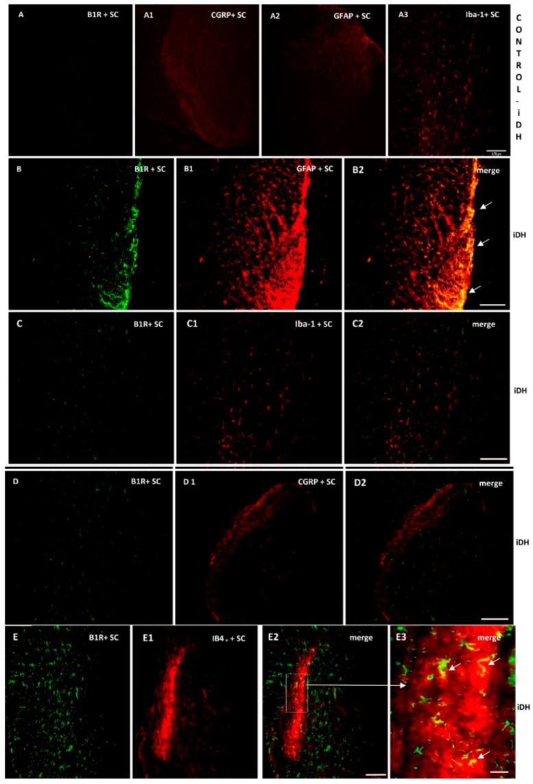
Kinins are mediators of pain and inflammation and evidence suggests that the inducible kinin B1 receptor (B1R) is involved in neuropathic pain (NP). This study investigates whether B1R and TRPV1 are colocalized on nociceptors and/or astrocytes to enable regulatory interaction either directly or through the cytokine pathway (IL-1β, TNF-α) in NP. Sprague Dawley rats were subjected to unilateral partial sciatic nerve ligation (PSNL) and treated from 14 to 21 days post-PSNL with antagonists of B1R (SSR240612, 10 mg·kg-1, i.p.) or TRPV1 (SB366791, 1 mg·kg-1, i.p.). The impact of these treatments was assessed on nociceptive behavior and mRNA expression of B1R, TRPV1, TNF-α, and IL-1β. Localization on primary sensory fibers, astrocytes, and microglia was determined by immunofluorescence in the lumbar spinal cord and dorsal root ganglion (DRG). Both antagonists suppressed PSNL-induced thermal hyperalgesia, but only SB366791 blunted mechanical and cold allodynia. SSR240612 reversed PSNL-induced enhanced protein and mRNA expression of B1R and TRPV1 mRNA levels in spinal cord while SB366791 further increased B1R mRNA/protein expression. B1R and TRPV1 were found in non-peptide sensory fibers and astrocytes, and colocalized in the spinal dorsal horn and DRG, notably with IL-1β on astrocytes. IL-1β mRNA further increased under B1R or TRPV1 antagonism. Data suggest that B1R and TRPV1 contribute to thermal hyperalgesia and play a distinctive role in allodynia associated with NP. Close interaction and reciprocal regulatory mechanism are suggested between B1R and TRPV1 on astrocytes and nociceptors in NP.

Keywords: B1 receptor; TRPV1; allodynia; astrocytes; bradykinin; cytokines; dorsal root ganglion; hyperalgesia; sensory fibers; spinal cord.


Conflict of interest statement

The authors declare no conflicts of interest. The funders had no role in the design of the study; in the collection, analyses, or interpretation of data; in the writing of the manuscript, or in the decision to publish the results.

Figures

Figure 1
Figure 1
Nociceptive behavior after treatment with kinin B1 receptor (B1R) antagonist. Effect of one-week daily administration of SSR240612 (10 mg·kg−1, i.p.) and its vehicle on mechanical allodynia (A), cold allodynia (B) and heat-hyperalgesia (C). Post-treatment values from days 1 to 7 are the average of the effect obtained during the first 2 h post-administration of the antagonist. Data represent the mean ± SEM of control (n = 7), vehicle-treated (n = 7) and SSR240612-treated (n = 8) groups. At some points, SEM are not visible because they overlap with the points. Control values are from the ipsilateral sham-operated side of untreated rats. Statistical analysis indicates * p < 0.05 vs. control and # p < 0.05 vs. vehicle-treated group, (F = 4.66, p < 0.001), for all three types of hypersensitivity.
Figure 2
Figure 2
Nociceptive behavior after treatment with transient receptor potential vanilloid 1 (TRPV1) antagonist. Effect of one-week daily administration of SB366791 (1 mg·kg−1, i.p.) and its vehicle on mechanical allodynia (A,B), cold allodynia (C,D), and heat-hyperalgesia (E). Time-course inhibitory effects of SB366791 on allodynia are given in B and D. Post-treatment values from days 1 to 7 in A and C represent the average of peak inhibition during the first 5–10 min post-administration. In E, PWL was still completely inhibited at 24 h post-treatment and therefore, the effect of the antagonist was similar before and after administration. Thus, time-course effect is not shown in E. Data represent the mean ± SEM of control (n = 7), vehicle-treated (n = 7) and SB366791-treated (n = 8) groups. At some points, SEM are not visible because they overlap with the points. Control values are from the ipsilateral sham-operated side of untreated rats. Statistical comparison to control is indicated by * p < 0.05, (F = 5.98, p <0.001), and to vehicle by # p < 0.05, (F = 5.98, p < 0.001), for all three types of hypersensitivity.
Figure 3
Figure 3
mRNA levels of B1R and TRPV1 in the spinal cord and dorsal root ganglion after treatment with SSR240612 and SB366791. mRNA levels of TRPV1 (white) and B1R (hatched) are shown in the contralateral and ipsilateral spinal cord (A,B) and dorsal root ganglion (C,D) of PSNL rats. Shown are the effects of one-week daily treatment with SSR240612 (10 mg·kg−1, A,C) and SB366791 (1.0 mg·kg−1, B,D). Values are fold-change ± SEM of control (n = 7), vehicle-treated (n = 7), SB366791-treated (n = 8) and SSR240612-treated (n = 8) groups. Statistical significance is indicated by * p < 0.05, (F = 4.66, p < 0.001) vs. control; # p < 0.05, (F = 4.66, p = 0.01) vs. vehicle; and + p < 0.05, (F = 4.66, p < 0.001) ipsilateral vehicle vs. contralateral vehicle. Each gene was compared to its own control for statistical analysis, yet only one control (black) is depicted in graphs (fold = 1).
Figure 4
Figure 4
B1R protein expression in the ipsilateral dorsal horn of the spinal cord. Images represent digital microphotographs of B1R protein expression in the ipsilateral dorsal horn of partial sciatic nerve ligation (PSNL) rats revealed by color-based (DAB chromogen) immuno-enzymatic (biotin-streptavidin HRP) technique. Quantitative optical density values (on a scale of 1.0) are given for B1R in control, vehicle-treated, SB366791 and SSR240612-treated rats. Values are the mean ± SEM of 5 rats per group (each value is the mean of 5 sections per rat). Significance is indicated by * p < 0.05 vs. control and # p < 0.05 vs. vehicle-treated group, (F = 3.95, p < 0.0001). Magnification is 40×.
Figure 5
Figure 5
Cellular distribution of B1R in the ipsilateral Dorsal Horn (iDH). Representative microphotographs by immunofluorescence of B1R (green, BE) relative to GFAP (red, B1), Iba-1 (red, C1), CGRP (red, D1) and IB4+ (red, E1) in the spinal cord (SC) of PSNL rats. Control spinal cord for B1R (A) expression exhibits very weak-to-no signal. Expression of CGRP (A1), GFAP (A2) and Iba-1 (A3) is moderate in the spinal cord of control animals. B1R immunolabelling is colocalized with astrocytes (arrows, GFAP, B2) and non-peptide fibers (arrows, IB4+, E2, E3), but not with microglia (Iba-1, C2) and peptide fibers (CGRP, D2) in the spinal dorsal horn of PSNL rats. E2 is enlarged in E3 as indicated by the white square and arrow. Scale bar = 120 µm (all panels, 10×), except 60 µm (E3, 20×).
Figure 6
Figure 6
Cellular distribution of TRPV1 in the ipsilateral Dorsal Horn (iDH). Representative microphotographs by immunofluorescence of TRPV1 (red, AD) relative to IB4+ (green, A1), CGRP (green, B1) GFAP (green, C1), Iba-1 (green, D1) in the spinal cord (SC) of PSNL rats. TRPV1 colocalizes weakly with peptide (CGRP, B2), and more abundantly with non-peptide fibers (IB4+, A2) and astroglia (GFAP, C2 C3), but not with microglia (D2). The same markers are shown in iDH of control rats. Scale bar = 120 µm (all panels, 10×), 60 µm (CC2 and DD2, 20×), 75 µm (C3, 60×).
Figure 7
Figure 7
Co-expression of B1R and TRPV1 in the ipsilateral Dorsal Horn (iDH). Representative microphotographs by immunofluorescence of B1R (green, A,B) and TRPV1 (red, A1,B1) in the spinal cord (SC) of PSNL rats. Images A2, B2 and B3 reveal a co-expression of B1R and TRPV1 (white arrows) at superficial layers of the dorsal spinal cord. Scale bar = 120 µm (AA2, 10×), 60 µm (BB2, 20×) and 50 µm (B3, 40×).
Figure 8
Figure 8
Co-expression of B1R with TRPV1 or GFAP in the ipsilateral lumbar DRG (iDRG). Representative microphotographs by immunofluorescence of B1R (green, B,C) and TRPV1 (red, B1,B2) and GFAP (red,C1) in the iDRG of PSNL rats. Control iDRG (A,A1,A2) exhibit no staining for B1R (A) and a moderate staining for both TRPV1 (A1) and GFAP (A2). B1R and TRPV1 are co-expressed in the DRG (B3). Enlarged DRG showing TRPV1 staining around the nucleus (arrow, B2). A punctual co-localization of B1R with GFAP (arrow, possibly issued from satellite cells) was found in the lumbar DRG (C2). Scale bar = 120 µm (AA2, 10×), 60 µm (other panels, 20×), 75 µm (B2, 60×).
Figure 9
Figure 9
Distribution of B1R and TRPV1 in relation with IL-1β in the ipsilateral Dorsal Horn (iDH). Representative microphotographs by immunofluorescence of B1R (green, A) and IL-1β (red, A1) in the spinal cord of PSNL rats. Image A2 shows colocalization between B1R and IL-1β (yellow spots). Likewise, TRPV1 (red, B) and IL-1β (green, B1) colocalize (B2) in the superficial layers of the spinal cord of PSNL rats. However, this colocalization is sparsely expressed. Scale bar = 120 µm. Magnification 10×.

References

    1. Van Hecke O., Austin S.K., Khan R.A., Smith B.H., Torrance N. Neuropathic pain in the general population: A systematic review of epidemiological studies. Pain. 2014;155:654–662. doi: 10.1016/j.pain.2013.11.013. - DOI - PubMed
    1. Woolf C.J. Dissecting out mechanisms responsible for peripheral neuropathic pain: Implications for diagnosis and therapy. Life Sci. 2004;74:2605–2610. doi: 10.1016/j.lfs.2004.01.003. - DOI - PubMed
    1. Couture R., Harrisson M., Vianna R.M., Cloutier F. Kinin receptors in pain and inflammation. Eur. J. Pharmacol. 2001;429:161–176. doi: 10.1016/S0014-2999(01)01318-8. - DOI - PubMed
    1. Calixto J.B., Medeiros R., Fernandes E.S., Ferreira J., Cabrini D.A., Campos M.M. Kinin B1 receptors: Key G-protein-coupled receptors and their role in inflammatory and painful processes. Br. J. Pharmacol. 2004;143:803–818. doi: 10.1038/sj.bjp.0706012. - DOI - PMC - PubMed
    1. Petcu M., Dias J.P., Ongali B., Thibault G., Neugebauer W., Couture R. Role of kinin B1 and B2 receptors in a rat model of neuropathic pain. Int. Immunopharmacol. 2008;8:188–196. doi: 10.1016/j.intimp.2007.09.009. - DOI - PubMed

LinkOut - more resources