. 2020 Jan 28;9(2):113.
doi: 10.3390/antiox9020113.

Potentilla rugulosa Nakai Extract Attenuates Bisphenol A-, S- and F-Induced ROS Production and Differentiation of 3T3-L1 Preadipocytes in the Absence of Dexamethasone

Affiliations

Potentilla rugulosa Nakai Extract Attenuates Bisphenol A-, S- and F-Induced ROS Production and Differentiation of 3T3-L1 Preadipocytes in the Absence of Dexamethasone

Sun-Il Choi et al. Antioxidants (Basel). .

Abstract

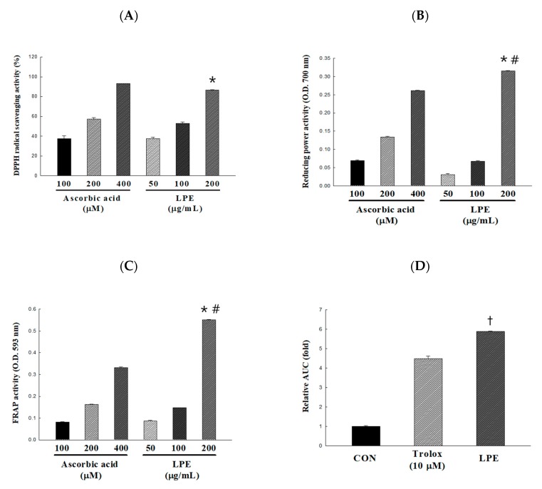
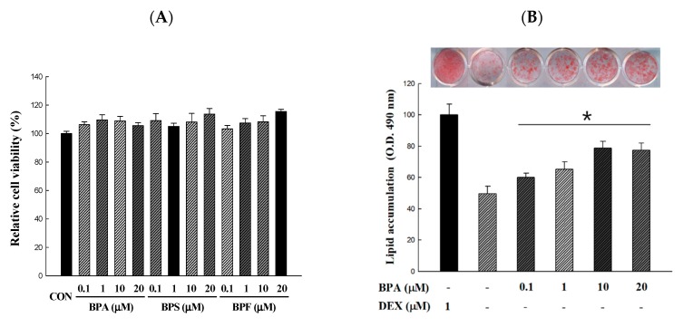
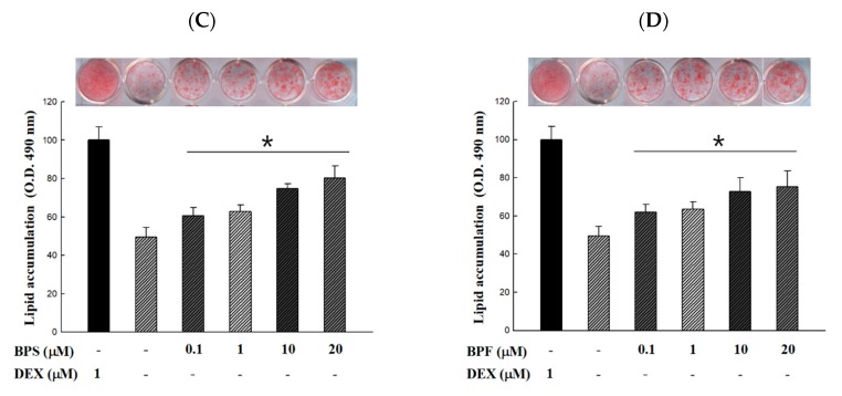
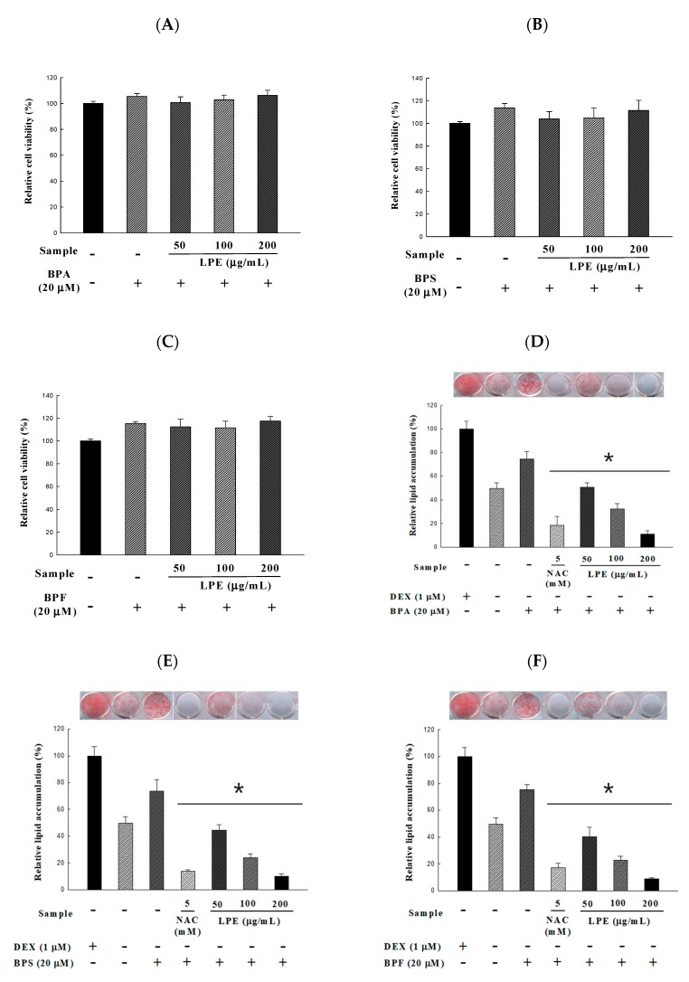
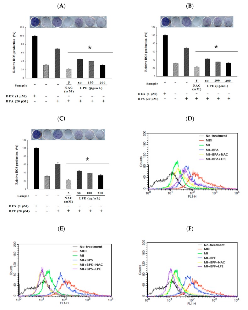
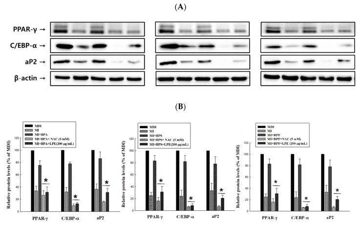
Endocrine disrupting chemicals (EDCs) disrupt the physiological metabolism, thus playing an important role in the development of obesity. EDCs, the so-called 'obesogens', might predispose some individuals to gain weight. This study investigated the effects of bisphenol A (BPA) and its alternatives (BPS and BPF) on adipocyte differentiation and the effects of the leaves of Potentilla rugulosa Nakai extract (LPE) as a functional food ingredient on obesogen-induced lipid production and adipogenesis in 3T3-L1 cells. The results showed that LPE has high total phenolic and flavonoid contents (77.58 ± 0.57 mg gallic acid equivalents (GAE)/g and 57.31 ± 1.72 mg quercetin equivalents (QE)/g, respectively). In addition, LPE exerted significant antioxidant effects in terms of DPPH radical scavenging activity, reducing power, ferric-ion reducing antioxidant power, and oxygen radical absorbance capacity. BPA, BPS, and BPF increased lipid accumulation, protein expressions of adipogenic transcription factors (PPAR-γ, C/EBP-α, and aP2), and reactive oxygen species (ROS) production in 3T3-L1 cells. However, LPE suppressed the BPA-, BPS-, and BPF-induced effects on adipogenesis. Therefore, LPE has potential as a functional food supplement that can prevent bisphenol-induced lipid metabolism disorders.

Keywords: Lipid metabolism disorders; Potentilla rugulosa Nakai; ROS production; bisphenol A; endocrine disrupting chemicals.


Conflict of interest statement

The authors declare no conflict of interest relevant to this study.

Figures

Figure 1
Figure 1
Antioxidant activity of the Potentilla rugulosa Nakai ethanolic extract (LPE) was established based on (A) DPPH radical-scavenging activity, (B) reducing power, (C) Ferric-ion reducing antioxidant power (FRAP) activity, and (D) areas under the curve fluorescence decay curve (AUC) value for ORAC. All values are presented as the mean ± SD. * p < 0.05 VS. 100 mg/mL of LPE; # p < 0.05 VS. 400 μM of ascorbic acid; p < 0.05 VS. 10 μM of trolox, according to one-way analysis of variance.
Figure 2
Figure 2
Cell viability and lipid accumulation from Bisphenol A (BPA), Bisphenol S (BPS), and Bisphenol F (BPF)-induced 3T3-L1 adipocyte. 3T3-L1 adipocytes were differentiated for 10 days with different concentrations of bisphenols. BPA, BPS, and BPF were treated 6 days before preadipocytes differentiation and maintained during cell differentiation. (A) Effect of BPA, BPS, and BPF on the viability was determined by an XTT assay and the absorbance at 450 nm was measured. (BD) The accumulated lipids were stained with Oil Red O reagent and the absorbance at 490 nm was measured. All values are presented as the mean ± SD. * p < 0.05 VS. MI treatment group, according to one-way analysis of variance.
Figure 2
Figure 2
Cell viability and lipid accumulation from Bisphenol A (BPA), Bisphenol S (BPS), and Bisphenol F (BPF)-induced 3T3-L1 adipocyte. 3T3-L1 adipocytes were differentiated for 10 days with different concentrations of bisphenols. BPA, BPS, and BPF were treated 6 days before preadipocytes differentiation and maintained during cell differentiation. (A) Effect of BPA, BPS, and BPF on the viability was determined by an XTT assay and the absorbance at 450 nm was measured. (BD) The accumulated lipids were stained with Oil Red O reagent and the absorbance at 490 nm was measured. All values are presented as the mean ± SD. * p < 0.05 VS. MI treatment group, according to one-way analysis of variance.
Figure 3
Figure 3
Effects of Potentilla rugulosa Nakai ethanolic extract (LPE) on BPA, BPS, and BPF-induced 3T3-L1 adipocyte in (AC) cell viability and (DF) lipid accumulation were determined by an XTT assay and ORO staining. 20 μM of BPA, BPS and BPF were treated 6 days before preadipocytes differentiation and maintained during cell differentiation. Various concentrations of LPE were dissolved in DMSO and treated on 3T3-L1 adipocyte (added on day 0 of differentiation) for 10 days. All values are presented as the mean ± SD. * p < 0.05 VS. MI treatment group, according to one-way analysis of variance.
Figure 4
Figure 4
Effects of LPE on BPA-, BPS-, and BPF-induced 3T3-L1 adipocyte in reactive oxygen species (ROS) were determined. After 10 days of 20 μM of bisphenol-induced adipocyte differentiation, ROS was measured by (AC) colorimetric analysis using NBT assay. Adipocyte were incubated with an NBT solution for 90 min, and the absorbance was measured at 593 nm. Another method of measuring ROS is (DF) fluorescence analysis using DCFDA assay. Adipocytes were incubated with 10 μM H2DCFDA for 30 min and the fluorescence was measured using flow cytometry. All values are presented as the mean ± SD. * p < 0.05 VS. MI treatment group, according to one-way analysis of variance.
Figure 5
Figure 5
Effects of LPE on the protein expressions of BPA-, BPS-, and BPF-induced adipogenic transcription factors (PPAR-γ: 53, 57kDa, C/EBP-α: 42 kDa and aP2: 15kDa) in 3T3-L1 adipocytes. (A) Total protein was obtained using a lysis buffer and quantified by loading, transfer, blocking, and appropriate antibody reactions. (B) Expression levels of adipogenic transcription factors were densitometrically normalized to those of β-actin and compared to the MDI control group as a percentage in each antibody. All values are presented as the mean ± SD. * p < 0.05 VS. BPA, BPS, or BPF treatment group, according to one-way analysis of variance.

Similar articles

Cited by

References

    1. Baillie-Hamilton P.F. Chemical toxins: A hypothesis to explain the global obesity epidemic. J. Altern. Complement. Med. 2002;8:185–192. doi: 10.1089/107555302317371479. - DOI - PubMed
    1. Neel B.A., Sargis R.M. The paradox of progress: environmental disruption of metabolism and the diabetes epidemic. Diabetes. 2011;60:1838–1848. doi: 10.2337/db11-0153. - DOI - PMC - PubMed
    1. Grün F., Blumberg B. Endocrine disrupters as obesogens. Mol. Cell Endocrinol. 2009;304:19–29. doi: 10.1016/j.mce.2009.02.018. - DOI - PMC - PubMed
    1. Grun F., Blumberg B. Perturbed nuclear receptor signaling by environmental obesogens as emerging factors in the obesity crisis. Rev. Endocr. Metab. Disord. 2007;8:161–171. doi: 10.1007/s11154-007-9049-x. - DOI - PubMed
    1. Legeay S., Faure S. Is bisphenol A an environmental obesogen? Fund Clin. Pharmacol. 2017;31:594–609. doi: 10.1111/fcp.12300. - DOI - PubMed