. 2020 Jan 28;21(3):843.
doi: 10.3390/ijms21030843.

Environmental Enrichment: Disentangling the Influence of Novelty, Social, and Physical Activity on Cerebral Amyloid Angiopathy in a Transgenic Mouse Model

Affiliations

Environmental Enrichment: Disentangling the Influence of Novelty, Social, and Physical Activity on Cerebral Amyloid Angiopathy in a Transgenic Mouse Model

Lisa S Robison et al. Int J Mol Sci. .

Abstract

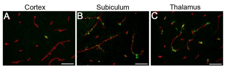
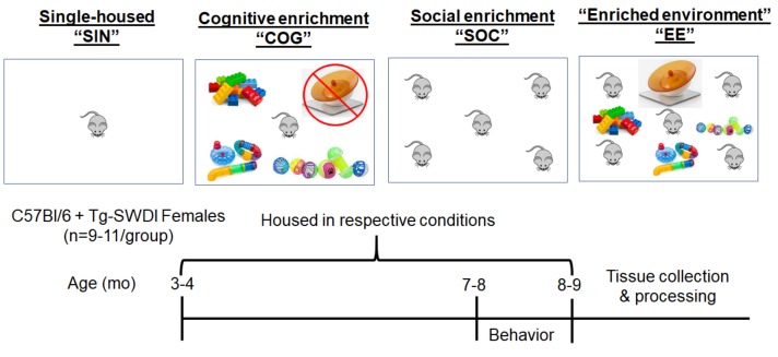
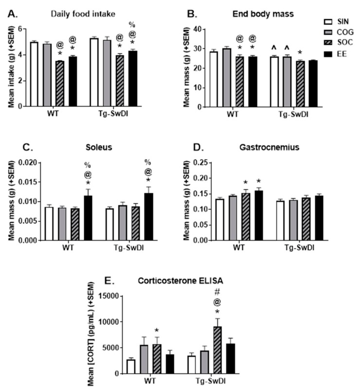
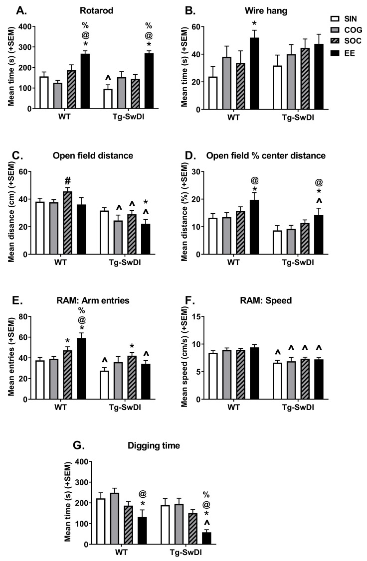
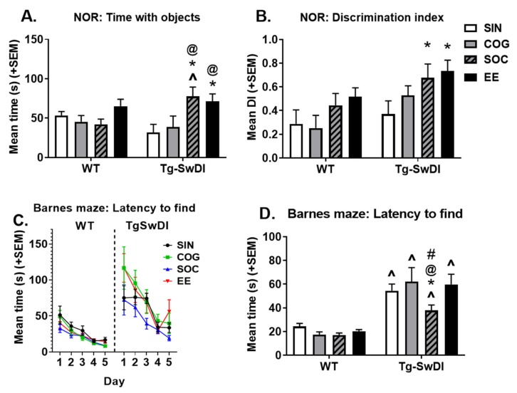
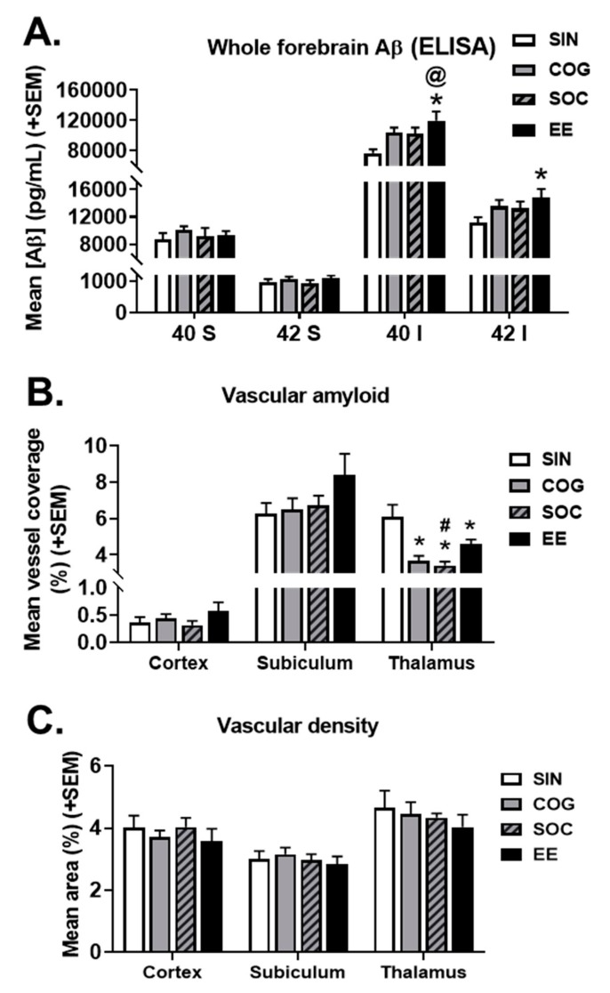
Cerebral amyloid angiopathy (CAA) is the deposition of amyloid protein in the cerebral vasculature, a common feature in both aging and Alzheimer's disease (AD). However, the effects of environmental factors, particularly cognitive stimulation, social stimulation, and physical activity, on CAA pathology are poorly understood. These factors, delivered in the form of the environmental enrichment (EE) paradigm in rodents, have been shown to have beneficial effects on the brain and behavior in healthy aging and AD models. However, the relative importance of these subcomponents on CAA pathology has not been investigated. Therefore, we assessed the effects of EE, social enrichment (SOC), and cognitive enrichment (COG) compared to a control group that was single housed without enrichment (SIN) from 4 to 8 months of age in wild-type mice (WT) and Tg-SwDI mice, a transgenic mouse model of CAA that exhibits cognitive/behavioral deficits. The results show that individual facets of enrichment can affect an animal model of CAA, though the SOC and combined EE conditions are generally the most effective at producing physiological, cognitive/behavioral, and neuropathological changes, adding to a growing literature supporting the benefits of lifestyle interventions.

Keywords: Alzheimer’s disease; cerebral amyloid angiopathy; enriched environment; exercise; reserve; resilience.


Conflict of interest statement

The authors declare no conflict of interest.

Figures

Figure 1
Figure 1
Cerebral microvascular amyloid accumulation in Tg-SwDI mice. Representative brain sections from 9-month-old Tg-SwDI mice stained with thioflavin S to identify fibrillar microvascular amyloid (green) and immunolabeled with an antibody to collagen IV to identify cerebral blood vessels (red). Microvascular amyloid deposits are observed in the cortex (A), subiculum (B), and thalamus (C). Scale bars = 50 µm.
Figure 2
Figure 2
Overview of housing conditions and timeline of the experiment. At ~3–4 months of age, C57Bl/6 and Tg-SwDI female mice were randomly assigned to one of four housing conditions: SIN (single housed), COG (single housed with cognitively stimulating toys switched out twice weekly), SOC (group housed), and EE (group housed mice with toys and running wheel). Following 4 months in the respective housing conditions, mice underwent a battery of behavior tests, and tissue was collected (end age = ~8–9 months). Within housing conditions, access to a running wheel is indicated by the presence of an orange exercise saucer. Cognitive enrichment is indicated by the presence of multi-colored blocks, balls, and tunnels.
Figure 3
Figure 3
Physiological measures. (A) Mean daily food intake over the course of the 4-month intervention period. Generally, Tg-SwDI mice ate more than WT mice, while SOC and EE housing attenuated food intake in WT and Tg-SwDI mice. (B) Body weight at the end of the experiment. Generally, the Tg-SwDI mice weighed less than the WT mice. SOC (both WT and Tg-SwDI), and EE (WT only) reduced body weight. (C) Soleus mass was increased by EE housing in both WT and Tg-SwDI mice. (D) Overall, Tg-SwDI mice tended to have a smaller gastrocnemius. There were trends of SOC and EE mice of both genotypes having a larger gastrocnemius, but this was only significant in WT mice. (E) Corticosterone levels measured by ELISA. * p < 0.05 vs. SIN of the same genotype, @ p < 0.05 vs. COG of the same genotype, % p < 0.05 vs. SOC of the same genotype, # p < 0.05 vs. EE of the same genotype, ^ p < 0.05 vs. WT in the same housing condition.
Figure 4
Figure 4
Activity and temperament measures. (A) Rotarod performance measured by time spent on rod (average of best two of three trials), indicative of balance and motor coordination. Under SIN conditions, Tg-SwDI mice were impaired in this task, while EE housing improved performance in both WT and Tg-SwDI mice. (B) Wire hang performance measured by time spent hanging onto a wire, indicative of forelimb strength. EE housing improved performance, but this was only significant in WT mice. (C) General activity levels, as measured by the distance traveled in the open field. SOC housing increased activity in WT mice. Overall, Tg-SwDI mice were hypoactive and this was exacerbated in EE mice. (D) Anxiety-like behavior, as measured by the percent of distance traveled in the center of the open field arena. Overall, Tg-SwDI mice exhibited increased anxiety-like behavior (less center activity), while anxiety was reduced by EE housing in both WT and Tg-SwDI mice. E and F) Exploratory behavior, as measured by the number of arm entries (E) and speed of travel (F) in the radial arm maze task. Generally, Tg-SwDI mice exhibited lower levels of exploration, and the number of arm entries was increased in SOC mice. In WT mice, SOC and EE housing increased the number of arm entries as well. (G) Time spent digging in a marble-burying task. Generally, Tg-SwDI mice exhibited lower levels of digging behavior, while EE housing attenuated digging behavior in both WT and Tg-SwDI mice. * p < 0.05 vs. SIN of the same genotype, @ p < 0.05 vs. COG of the same genotype, % p < 0.05 vs. SOC of the same genotype, # p < 0.05 vs. EE of the same genotype, ^ p < 0.05 vs. WT in the same housing condition.
Figure 5
Figure 5
Cognitive testing. In Tg-SwDI mice, SOC and EE housing increased the amount of time spent with objects (A), and the discrimination index (B) in the novel object recognition (NOR) task. (C) Barnes maze performance, as measured by the latency to find the escape hole over time. (D) Barnes maze performance, as measured by the average latency to find the escape hole on days 2–5. Tg-SwDI mice took longer to find the escape hole compared to WT mice in all housing conditions. In Tg-SwDI mice, SOC housing was associated with improved performance (shorter latency to find the escape hole). * p < 0.05 vs. SIN of the same genotype, @ p < 0.05 vs. COG of the same genotype, % p < 0.05 vs. SOC of the same genotype, # p < 0.05 vs. EE of the same genotype, ^ p < 0.05 vs. WT in the same housing condition.
Figure 6
Figure 6
Effects of the housing condition on the accumulation of Aβ in Tg-SwDI mice. (A) Tg-SwDI mice housed in EE conditions had increased insoluble Aβ40 and Aβ42) levels, as measured by ELISA of whole forebrain homogenates. (B) Tg-SwDI mice in all enrichment conditions had less vascular amyloid in the thalamus compared to single-housed mice. (C) The housing condition did not affect the cerebral vascular density in any of the three brain regions measured. * p < 0.05 vs. SIN of the same genotype, @ p < 0.05 vs. COG of the same genotype, # p < 0.05 vs. EE of the same genotype.

References

    1. Yamada M. Risk factors for cerebral amyloid angiopathy in the elderly. Ann. New York Acad. Sci. 2002;977:37–44. doi: 10.1111/j.1749-6632.2002.tb04797.x. - DOI - PubMed
    1. Kinnecom C., Lev M., Wendell L., Smith E., Rosand J., Frosch M., Greenberg S. Course of cerebral amyloid angiopathy–related inflammation. Neurology. 2007;68:1411–1416. doi: 10.1212/01.wnl.0000260066.98681.2e. - DOI - PubMed
    1. Suter O.-C., Sunthorn T., Kraftsik R., Straubel J., Darekar P., Khalili K., Miklossy J. Cerebral hypoperfusion generates cortical watershed microinfarcts in Alzheimer disease. Stroke. 2002;33:1986–1992. doi: 10.1161/01.STR.0000024523.82311.77. - DOI - PubMed
    1. Holland C.M., Smith E.E., Csapo I., Gurol M.E., Brylka D.A., Killiany R.J., Blacker D., Albert M.S., Guttmann C.R., Greenberg S.M. Spatial distribution of white-matter hyperintensities in Alzheimer disease, cerebral amyloid angiopathy, and healthy aging. Stroke. 2008;39:1127–1133. doi: 10.1161/STROKEAHA.107.497438. - DOI - PMC - PubMed
    1. Yoshita M., Fletcher E., Harvey D., Ortega M., Martinez O., Mungas D., Reed B., DeCarli C. Extent and distribution of white matter hyperintensities in normal aging, MCI, and AD. Neurology. 2006;67:2192–2198. doi: 10.1212/01.wnl.0000249119.95747.1f. - DOI - PMC - PubMed

Substances