. 2020 Jan 29;9(2):365.
doi: 10.3390/jcm9020365.

Shoulder Muscle Strength and Neuromuscular Activation 2 Years after Reverse Shoulder Prosthesis-An Experimental Case Control Study

Affiliations

Shoulder Muscle Strength and Neuromuscular Activation 2 Years after Reverse Shoulder Prosthesis-An Experimental Case Control Study

Anna Rienmüller et al. J Clin Med. .

Abstract

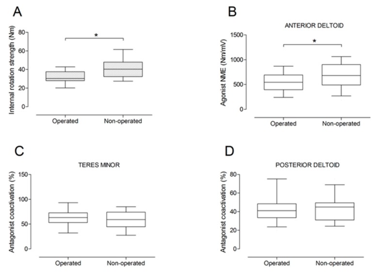
Although reverse shoulder arthroplasty (RSA) has shown successful postoperative outcomes, little is known about compensatory activation patterns of remaining shoulder muscles following RSA. The purpose of this experimental case control series was to investigate shoulder muscle strength and neuromuscular activation of deltoid and teres minor muscles 2 years after RSA. Humerus lengthening, center-of-rotation medialization, maximal voluntary strength, and electromyographic (EMG) activity were compared between the operated and the non-operated side of 13 patients (mean age: 73 years). Shoulder muscle strength was significantly lower on the operated side for external rotation (-54%), internal rotation (-20%), and adduction (-13%). Agonist deltoid EMG activity was lower on the operated side for shoulder flexion, extension, and internal and external rotation (p < 0.05). Antagonist deltoid coactivation was higher on the operated side for external rotation (p < 0.001). Large correlation coefficients were observed between shoulder adductor strength asymmetry and both center-of-rotation medialization (r = -0.73) and humerus lengthening (r = 0.71). Shoulder abduction strength and neuromuscular activation were well preserved 2 years after RSA, while persistent strength and activation deficits were observed for shoulder adduction and internal and external rotation. Additional studies are required to elucidate shoulder neuromuscular activation patterns before and after RSA to support decision making for surgical, implant design, and rehabilitation choices.

Keywords: deltoid muscle; electromyography; humerus lengthening; muscle strength; neuromuscular activation; reverse shoulder arthroplasty.


Conflict of interest statement

The authors declare no conflict of interest.

Figures

Figure 1
Figure 1
Anteroposterior radiographs of the same patient pre- and post-operatively are shown. Evaluation of center-of-rotation (COR) medialization was performed according to the protocol of Greiner et al. [5]. Pre-operatively (a), the distance “mo” was measured between the center of the circle involving the articular surface (COR) and the vertical line from the outer border of the acromion. Post-operatively (b), the distance “m” was measured between the center of the base plate (COR) and the vertical line from the outer border of the acromion. COR medialization was calculated as the difference between the distances “mo” and “m” as follows: ∆m = mo − m. mo > m was found in all subjects (12.8 mm for this representative patient).
Figure 2
Figure 2
Measurement of humerus lengthening according to the protocol of Greiner et al. [5] and Lädermann et al. [10]. The humeral shaft axis was drawn, and the distance between the two lines “a” (subacromial line) and “e” (inter-epicondylar line) was measured as the humerus length for the operated (lo) (a) and non-operated side (lc) (b). The difference between the operated and non-operated humerus length (∆l = lo − lc) was considered as humerus lengthening if lo > lc. (8 mm for this representative patient).
Figure 3
Figure 3
Overview of shoulder actions with respective agonist and antagonist muscles evaluated with surface electromyographic (EMG) activity in this study.
Figure 4
Figure 4
Box and whisker plots of shoulder abduction strength (A) and agonist neuromuscular efficiency (NME) of lateral deltoid (B) and anterior deltoid (C) muscles by side. The horizontal line in the box represents the median, the height of the box represents the interquartile range, and the distance between the opposite ends of the whisker represents the 10th–90th percentile.
Figure 5
Figure 5
Box and whisker plots of shoulder adduction strength (A) and antagonist coactivation of lateral deltoid (B) and anterior deltoid (C) muscles by side. The horizontal line in the box represents the median, the height of the box represents the interquartile range, and the distance between the opposite ends of the whisker represents the 10th–90th percentile. *Operated < non-operated (p < 0.05).
Figure 6
Figure 6
Box and whisker plots of shoulder flexion strength (A), agonist neuromuscular efficiency (NME) of anterior deltoid (B) and lateral deltoid (C) muscles, and antagonist coactivation of posterior deltoid muscle (D) by side. The horizontal line in the box represents the median, the height of the box represents the interquartile range, and the distance between the opposite ends of the whisker represents the 10th–90th percentile. *Operated < non-operated (p < 0.05).
Figure 7
Figure 7
Box and whisker plots of shoulder extension strength (A), agonist neuromuscular efficiency (NME) of posterior deltoid muscle (B), antagonist coactivation of anterior deltoid (C) and lateral deltoid (D) muscles by side. The horizontal line in the box represents the median, the height of the box represents the interquartile range, and the distance between the opposite ends of the whisker represents the 10th–90th percentile. *Operated < non-operated (p < 0.05).
Figure 8
Figure 8
Box and whisker plots of shoulder internal rotation strength (A), agonist neuromuscular efficiency (NME) of anterior deltoid muscle (B), antagonist coactivation of teres minor (C) and posterior deltoid (D) muscles by side. The horizontal line in the box represents the median, the height of the box represents the interquartile range, and the distance between the opposite ends of the whisker represents the 10th–90th percentile. *Operated < non-operated (p < 0.05).
Figure 9
Figure 9
Box and whisker plots of shoulder external rotation strength (A), agonist neuromuscular efficiency (NME) of teres minor (B) and posterior deltoid (C) muscles, and antagonist coactivation of anterior deltoid muscle (D). The horizontal line in the box represents the median, the height of the box represents the interquartile range, and the distance between the opposite ends of the whisker represents the 10th–90th percentile. * Operated ≠ non-operated (p < 0.05).
Figure 10
Figure 10
Correlations between shoulder adductor strength asymmetry and COR medialization (A) or humerus lengthening (B).

References

    1. Grammont P.M., Baulot E. Delta shoulder prosthesis for rotator cuff rupture. Orthopedics. 1993;16:65–68. doi: 10.3928/0147-7447-19930101-11. - DOI - PubMed
    1. Boileau P., Watkinson D.J., Hatzidakis A.M., Balg F. Grammont reverse prosthesis: Design, rationale, and biomechanics. J. Shoulder Elbow Surg. 2005;14:147S–161S. doi: 10.1016/j.jse.2004.10.006. - DOI - PubMed
    1. Jobin C.M., Brown G.D., Bahu M.J., Gardner T.R., Bigliani L.U., Levine W.N., Ahmad C.S. Reverse total shoulder arthroplasty for cuff tear arthropathy: the clinical effect of deltoid lengthening and center of rotation medialization. J. Shoulder Elbow Surg. 2012;21:1269–1277. doi: 10.1016/j.jse.2011.08.049. - DOI - PubMed
    1. Cuff D., Clark R., Pupello D., Frankle M. Reverse shoulder arthroplasty for the treatment of rotator cuff deficiency: A concise follow-up, at a minimum of five years, of a previous report. J. Bone Joint Surg. Am. 2012;94:1996–2000. doi: 10.2106/JBJS.K.01206. - DOI - PubMed
    1. Greiner SH., Back D.A., Herrmann S., Perka C., Asbach P. Degenerative changes of the deltoid muscle have impact on clinical outcome after reversed total shoulder arthroplasty. Arch. Orthop. Trauma Surg. 2010;130:177–183. doi: 10.1007/s00402-009-1001-y. - DOI - PubMed

LinkOut - more resources