. 2020 Jan 29;12(2):152.
doi: 10.3390/v12020152.

Characteristics of Human OAS1 Isoform Proteins

Affiliations

Characteristics of Human OAS1 Isoform Proteins

Han Di et al. Viruses. .

Abstract

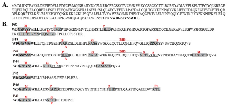
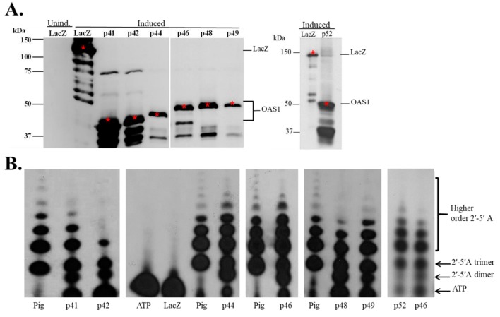
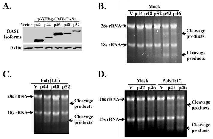
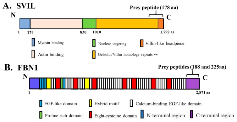
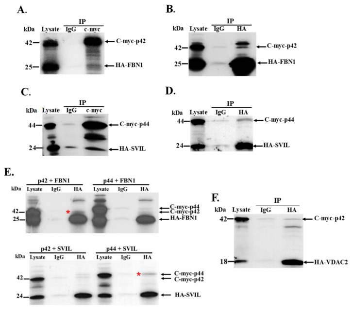
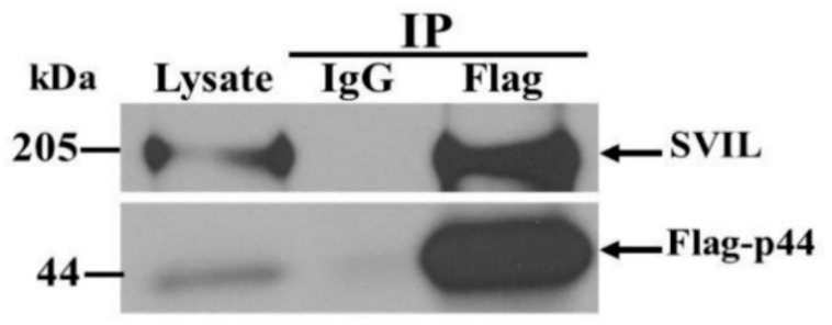
The human OAS1 (hOAS1) gene produces multiple possible isoforms due to alternative splicing events and sequence variation among individuals, some of which affect splicing. The unique C-terminal sequences of the hOAS1 isoforms could differentially affect synthetase activity, protein stability, protein partner interactions and/or cellular localization. Recombinant p41, p42, p44, p46, p48, p49 and p52 hOAS1 isoform proteins expressed in bacteria were each able to synthesize trimer and higher order 2'-5' linked oligoadenylates in vitro in response to poly(I:C). The p42, p44, p46, p48 and p52 isoform proteins were each able to induce RNase-mediated rRNA cleavage in response to poly(I:C) when overexpressed in HEK293 cells. The expressed levels of the p42 and p46 isoform proteins were higher than those of the other isoforms, suggesting increased stability in mammalian cells. In a yeast two-hybrid screen, Fibrillin1 (FBN1) was identified as a binding partner for hOAS1 p42 isoform, and Supervillin (SVIL) as a binding partner for the p44 isoform. The p44-SVIL interaction was supported by co-immunoprecipitation data from mammalian cells. The data suggest that the unique C-terminal regions of hOAS1 isoforms may mediate the recruitment of different partners, alternative functional capacities and/or different cellular localization.

Keywords: 2-5A; Fibrillin1; Supervillin; human OAS1 isoforms; rRNA cleavage.


Conflict of interest statement

The authors declare no conflicts of interest.

Figures

Figure 1
Figure 1
The unique C-terminal amino acid sequences of seven hOAS1 isoform proteins. (A) The common N-terminal sequence shared by all of the hOAS1 isoforms. The bolded C terminal sequence is repeated in the bottom panel. (B) The unique C-terminal sequences of the isoforms. Predicted post-translational modification sites or domains are indicated by red letters. M, N-myristoylation site; P, various predicted phosphorylation sites; G, N-linked glycosylation site; A, amidation site; Pr, prenylation site; BH3, BH3 domain. Post translational modification sites were predicted using PROSITE (https://prosite.expasy.org).
Figure 2
Figure 2
Bacterial expression of hOAS1 isoform proteins and analysis of their ability to produce 2-5A. (A) Seven hOAS1 isoform fusion proteins and LacZ protein were expressed individually in bacteria. After isopropyl β-d-1-thiogalacto-pyranoside (IPTG) (1 mM) induction, bacteria were lysed and proteins in the crude bacterial lysates were separated by 10% sodium dodecyl sulfate-polyacrylamide gel electrophoresis (SDS-PAGE), transferred to a nitrocellulose membrane and immunoblotted with anti-V5 antibody. All of the OAS1 isoforms except for p52 were run on the same gel. A lysate from uninduced bacteria transformed with a plasmid containing the LacZ gene was used as a negative control. The full-length proteins are indicated by a red asterisk. OAS1 degradation bands were detected on the gel below the full-length proteins. (B) After protein purification on a Talon metal affinity column, each human OAS1 isoform (22 μL) was incubated with α32P-adenosine triphosphate (ATP) and poly(I:C) for 18 h at 30 °C. LacZ protein plus α32P-ATP (Lane 5) and α32P-ATP alone (no protein) (Lane 4) were used as negative controls. Purified Escherichia coli (E. coli)-expressed pig OAS1 (0.5 μg) was used as a positive control. Two µL of each reaction were electrophoresed on a 20% polyacrylamide/8M urea denaturing gel and radiolabeled 2-5A was visualized by autoradiography. Separate gels used to analyze the 2-5A are indicated by spaces.
Figure 3
Figure 3
Analysis of RNase L cleavage in cells overexpressing individual hOAS1 isoforms in response to poly(I:C). HEK293 cells were transfected with 1 µg of empty vector DNA, or of one of the hOAS1 isoforms cloned into the p3xFlag-CMV plasmid for 24 h. (A) Western blot analysis of the expression level of each OAS1 isoform, using anti-Flag and anti-β actin antibodies. HEK293 cells overexpressing each OAS1 isoform (1 µg of plasmid DNA) for 24 h were either mock transfected (B), or transfected with poly(I:C) for 6 h (C). Total intracellular RNA was extracted and separated on a denaturing agarose gel and stained with ethidium bromide. (D) HEK293 cells were transfected with 0.5 µg p42 or p46 plasmid DNA for 9 h, followed by either mock transfection, or transfection with poly(I:C) for 6 h. Total intracellular RNA was extracted and separated on a denaturing agarose gel and stained with ethidium bromide. The 18S/28S rRNA and their cleavage products are indicated by arrows. V: empty vector control.
Figure 4
Figure 4
(A) Diagram of the domain structure of the full-length SVIL protein. The domains are color-coded, and the location of the 178 aa prey peptide (aa 1615–1792) is indicated by a bracket. The amino acid numbers are indicated below the diagram. (B) Diagram of the domain structure of the full-length FBN1 protein. The domains are color-coded, and the location of the two overlapping prey peptides, 188 aa (aa 2684–2871) and 225 aa (aa 2647–2871), are indicated by a bracket. EGF: epidermal growth factor.
Figure 5
Figure 5
Analysis of the interaction between human OAS1 p42 and p44 isoforms and their respective binding partners in vitro. Wheat germ extract was used for the in vitro transcription and translation of c-myc tagged full length human OAS1 isoforms and of HA-tagged FBN1 (188 aa), SVIL or voltage-dependent anion channel (VDAC) peptides in the presence of [35S]-methionine. Reciprocal pull-down assays were performed with anti-HA and anti-c-myc antibodies. Non-specific IgG was used as the negative control. The protein complexes were resolved by SDS-PAGE and detected by autoradiography. (A) HA-FBN1 peptide and full-length c-myc-OAS1 p42 immunoprecipitated with anti-c-myc antibody. (B) C-myc-OAS1 p42 and HA-FBN1 peptide immunoprecipitated with anti-HA antibody. (C) HA-SVIL peptide and c-myc-OAS1 p44 immunoprecipitated with anti-c-myc antibody. (D) C-myc-OAS1 p44 and HA-SVIL peptide immunoprecipitated with anti-HA antibody (E) (top panel) Either C-myc-OAS1 p42 or p44 and HA-FBN1 peptide immunoprecipitated with anti-HA antibody and (bottom panel) either C-myc-OAS1 p42 or p44 and HA-SVIL peptide immunoprecipitated with anti-HA antibody. The hOAS1 isoform specifically immunoprecipitated is indicated with a red asterisk. (F) C-myc-OAS1 p42 and HA-VDAC2 peptide immunoprecipitated with anti-HA antibody. IgG: non-specific antibody control; IP: immunoprecipitation.
Figure 6
Figure 6
Interaction between the hOAS1 p44 isoform and endogenous full-length SVIL in A549 cell extracts. Human A549 cells were transiently transfected with plasmid DNA expressing the 3xFlag-tagged full-length hOAS1 p44 isoform. At 48 h post transfection, cell lysates were collected and incubated with anti-Flag antibody-conjugated agarose beads. Non-specific IgG conjugated agarose beads were used as the negative control. After co-immunoprecipitation, protein complexes were resolved by SDS-PAGE, transferred to a nitrocellulose membrane and detected by anti-Flag and anti-SVIL antibodies.

References

    1. Kristiansen H., Gad H.H., Eskildsen-Larsen S., Despres P., Hartmann R. The oligoadenylate synthetase family: An ancient protein family with multiple antiviral activities. J. Interferon Cytokine Res. Off. J. Int. Soc. Interferon Cytokine Res. 2011;31:41–47. doi: 10.1089/jir.2010.0107. - DOI - PubMed
    1. Kjaer K.H., Poulsen J.B., Reintamm T., Saby E., Martensen P.M., Kelve M., Justesen J. Evolution of the 2’–5’-oligoadenylate synthetase family in eukaryotes and bacteria. J. Mol. Evol. 2009;69:612–624. doi: 10.1007/s00239-009-9299-1. - DOI - PubMed
    1. Rebouillat D., Marie I., Hovanessian A.G. Molecular cloning and characterization of two related and interferon-induced 56-kDa and 30-kDa proteins highly similar to 2′–5′ oligoadenylate synthetase. Eur. J. Biochem. 1998;257:319–330. doi: 10.1046/j.1432-1327.1998.2570319.x. - DOI - PubMed
    1. Marie I., Hovanessian A.G. The 69-kDa 2-5A synthetase is composed of two homologous and adjacent functional domains. J. Biol. Chem. 1992;267:9933–9939. - PubMed
    1. Hartmann R., Olsen H.S., Widder S., Jorgensen R., Justesen J. p59OASL, a 2′–5′ oligoadenylate synthetase like protein: A novel human gene related to the 2′–5′ oligoadenylate synthetase family. Nucleic Acids Res. 1998;26:4121–4128. doi: 10.1093/nar/26.18.4121. - DOI - PMC - PubMed

Publication types