. 2020 Jan 29;12(2):312.
doi: 10.3390/cancers12020312.

Development of a Precision Medicine Workflow in Hematological Cancers, Aalborg University Hospital, Denmark

Affiliations

Development of a Precision Medicine Workflow in Hematological Cancers, Aalborg University Hospital, Denmark

Julie S Bødker et al. Cancers (Basel). .

Abstract

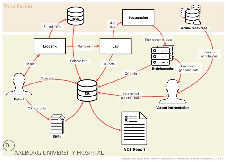
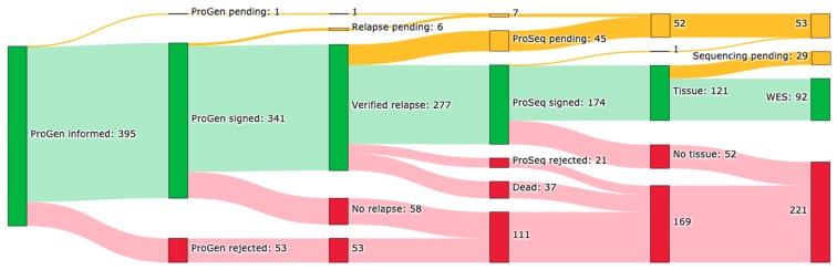
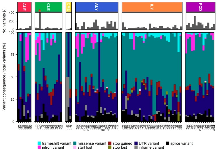
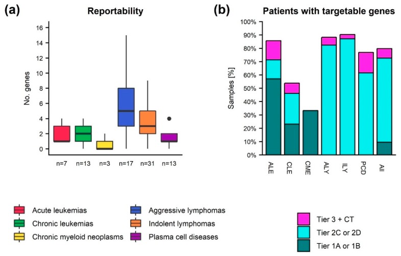
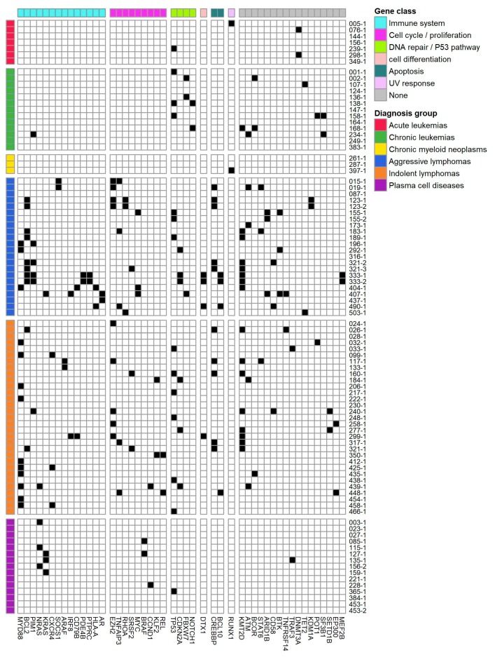
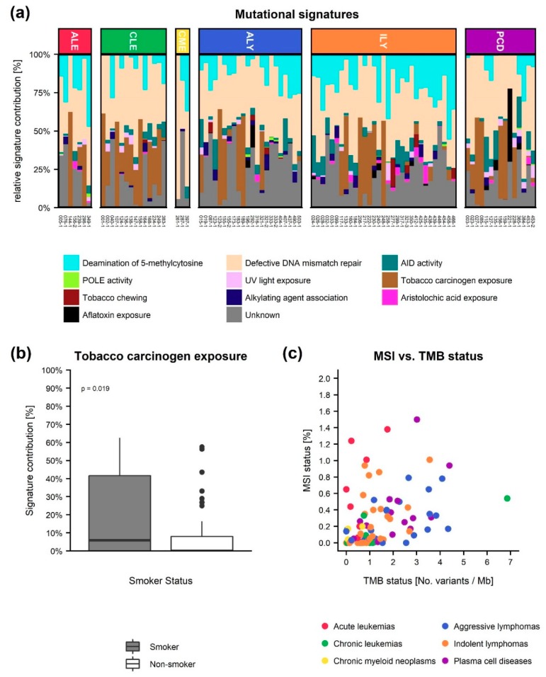
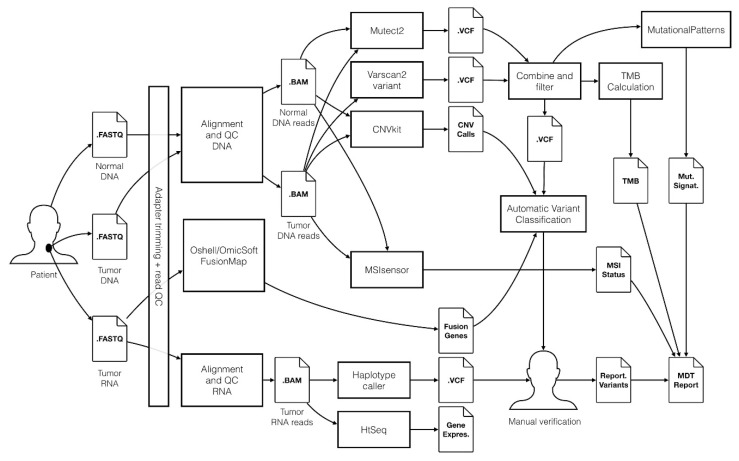
Within recent years, many precision cancer medicine initiatives have been developed. Most of these have focused on solid cancers, while the potential of precision medicine for patients with hematological malignancies, especially in the relapse situation, are less elucidated. Here, we present a demographic unbiased and observational prospective study at Aalborg University Hospital Denmark, referral site for 10% of the Danish population. We developed a hematological precision medicine workflow based on sequencing analysis of whole exome tumor DNA and RNA. All steps involved are outlined in detail, illustrating how the developed workflow can provide relevant molecular information to multidisciplinary teams. A group of 174 hematological patients with progressive disease or relapse was included in a non-interventional and population-based study, of which 92 patient samples were sequenced. Based on analysis of small nucleotide variants, copy number variants, and fusion transcripts, we found variants with potential and strong clinical relevance in 62% and 9.5% of the patients, respectively. The most frequently mutated genes in individual disease entities were in concordance with previous studies. We did not find tumor mutational burden or micro satellite instability to be informative in our hematologic patient cohort.

Keywords: bioinformatics workflow; hematology; next generation sequencing; precision medicine; somatic cancer variants; variant interpretation.


Conflict of interest statement

The authors declare no conflict of interest.

Figures

Figure A1
Figure A1
Laboratory Information Management. An overview of the data flow in the ProGen and ProSeq studies. Patient consent is stored in a REDCap database (DB), and clinical data of the patient was from the electronic health records (EHRs) are transferred into the REDCap database. Donated tissue samples are handled and stored in the biobank, and sample information is subsequently placed in the national RBGB database, and automatically imported into the RedCap database, minimizing the time consumption for patient registration. In the laboratory (lab), DNA and RNA is purified from tissue samples or from sorted malignant cells and subsequently sequenced. Raw genomic data is processed using the bioinformatics workflow, and the resulting genomic profile is annotated using online cancer database resources. Following, quality control (QC) data from the Lab and the Bioinformatics analysis is recorded in the REDcap database. Variant interpretation is performed by trained molecular biologists and the resulting interpreted genomic data is transferred to REDCap. Finally, a report for multidisciplinary team conferences (MDT Report) can be generated for each patient.
Figure 1
Figure 1
Inclusion overview. Number of patient cases included in the ProGen and ProSeq studies by 31 January 2019. Patient’s relapse status, presence of correct tumor tissue for analysis, and sample status is given. Color codes: green: yes, yellow: pending, and red: no.
Figure 2
Figure 2
Histograms of sample sequencing depth. (a) Average target coverage of normal and tumor Whole Exome Sequencing (WES) samples. (b) Total mapped paired end RNAseq reads. Dashed lines indicate the minimum sample depth requirements.
Figure 3
Figure 3
Small nucleotide variant (SNV) detection and filtering. Retained variants per patient grouped by type and divided into the six disease groups; ALE: Acute leukemia, CLE: Chronic leukemia, CME: Chronic Myeloid Neoplasms, ALY: Aggressive Lymphomas, ILY: Indolent Lymphomas, and PCD: Plasma Cell Diseases. * Sample contained 5061 variants and was not subjected to variant interpretation in Qiagen Clinical Insight Interpret.
Figure 4
Figure 4
Reportable somatic variants in hematological malignancies. (a) Number of genes with detected somatic variants reported per sample after manual verification. (b) Percentage of patients (grouped by diagnosis or overall) with at least one variant having strong clinical relevance (tier 1A and 1B), potential clinical relevance (tier 2C and 2D), or at most having unknown clinical significance, but with an associated clinical trial (tier 3 + CT). If multiple samples were sequenced for a patient, only the latest is represented in the figure. The single sample from a patient with CLL with more than 5000 retained variants was not subjected to variant interpretation. ALE: Acute leukemia, CLE: Chronic leukemia, CME: Chronic Myeloid Neoplasms, ALY: Aggressive Lymphomas, ILY: Indolent Lymphomas, PCD: Plasma Cell Diseases.
Figure 5
Figure 5
Occurrence of genes with clinically relevant alterations. The genomic landscape of distinct, clinically relevant gene alterations across various hematologic cancers if observed in more than one patient. Each row represents a patient sample. These are grouped by diagnosis group. Each column represents gene with clinical relevant alterations. Genes are organized by gene sets derived from MSigDB Collection2 (Version 6.2) [23,24,25].
Figure 6
Figure 6
Other potential clinically relevant genomic measurements. (a) The relative contribution of the COMSIC mutational signatures to each tumor sample grouped as either Deamination of 5−methylcytosine, defective DNA mismatch repair, activation-induced cytidine deaminase (AID) activity, DNA polymerase epsilon (POLE) activity, UV light exposure, tobacco carcinogen exposure, tobacco chewing, alkylating agent association, aristolochic acid exposure, or aflatoxin exposure according to their proposed aetiology. ALE: Acute leukemia, CLE: Chronic leukemia, CMNE: Chronic Myeloid Neoplasms, ALY: Aggressive Lymphomas, ILY: Indolent Lymphomas and PCD: Plasma Cell Diseases. (b) Comparison of the relative contribution of the tobacco carcinogen exposure signature in the smoker vs. non-smoker groups. (c) Micro satellite instability (MSI) status vs. tumor mutational burden (TMB) status.
Figure 7
Figure 7
Overview of the bioinformatics workflow for processing of WES and RNA sequencing data in the ProSeq study. The workflow is based on the Genome Analysis Tool Kit (GATK) framework, and Qiagen Clinical Insight Interpret is used to support the manual variant interpretation performed by molecular biologists. Genomic data results are stored in a precision cancer medicine database, potentially enabling results to be shared with other hospital organizations and the scientific community.

References

    1. Le Tourneau C., Delord J.P., Gonçalves A., Gavoille C., Dubot C., Isambert N., Campone M., Trédan O., Massiani M.A., Mauborgne C., et al. Molecularly targeted therapy based on tumour molecular profiling versus conventional therapy for advanced cancer (SHIVA): A multicentre, open-label, proof-of-concept, randomised, controlled phase 2 trial. Lancet Oncol. 2015;16:1324–1334. doi: 10.1016/S1470-2045(15)00188-6. - DOI - PubMed
    1. Conley B.A., Doroshow J.H. Molecular Analysis for Therapy Choice: NCI MATCH. Semin. Oncol. 2014;41:297–299. doi: 10.1053/j.seminoncol.2014.05.002. - DOI - PubMed
    1. Levit L.A., Kim E.S., McAneny B.L., Nadauld L.D., Levit K., Schenkel C., Schilsky R.L. Implementing Precision Medicine in Community-Based Oncology Programs: Three Models. J. Oncol. Pract. 2019;15:325–329. doi: 10.1200/JOP.18.00661. - DOI - PubMed
    1. Tyner J.W., Tognon C.E., Bottomly D., Wilmot B., Kurtz S.E., Savage S.L., Long N., Schultz A.R., Traer E., Abel M., et al. Functional genomic landscape of acute myeloid leukaemia. Nature. 2018 doi: 10.1038/s41586-018-0623-z. - DOI - PMC - PubMed
    1. Miao Y., Medeiros L.J., Li Y., Li J., Young K.H. Genetic alterations and their clinical implications in DLBCL. Nat. Rev. Clin. Oncol. 2019;16:634–652. doi: 10.1038/s41571-019-0225-1. - DOI - PubMed

LinkOut - more resources