Angiotensin-II-Evoked Ca2+ Entry in Murine Cardiac Fibroblasts Does Not Depend on TRPC Channels
- PMID: 32013125
- PMCID: PMC7072683
- DOI: 10.3390/cells9020322
Angiotensin-II-Evoked Ca2+ Entry in Murine Cardiac Fibroblasts Does Not Depend on TRPC Channels
Abstract
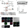
TRPC proteins form cation conducting channels regulated by different stimuli and are regulators of the cellular calcium homeostasis. TRPC are expressed in cardiac cells including cardiac fibroblasts (CFs) and have been implicated in the development of pathological cardiac remodeling including fibrosis. Using Ca2+ imaging and several compound TRPC knockout mouse lines we analyzed the involvement of TRPC proteins for the angiotensin II (AngII)-induced changes in Ca2+ homeostasis in CFs isolated from adult mice. Using qPCR we detected transcripts of all Trpc genes in CFs; Trpc1, Trpc3 and Trpc4 being the most abundant ones. We show that the AngII-induced Ca2+ entry but also Ca2+ release from intracellular stores are critically dependent on the density of CFs in culture and are inversely correlated with the expression of the myofibroblast marker α-smooth muscle actin. Our Ca2+ measurements depict that the AngII- and thrombin-induced Ca2+ transients, and the AngII-induced Ca2+ entry and Ca2+ release are not affected in CFs isolated from mice lacking all seven TRPC proteins (TRPC-hepta KO) compared to control cells. However, pre-incubation with GSK7975A (10 µM), which sufficiently inhibits CRAC channels in other cells, abolished AngII-induced Ca2+ entry. Consequently, we conclude the dispensability of the TRPC channels for the acute neurohumoral Ca2+ signaling evoked by AngII in isolated CFs and suggest the contribution of members of the Orai channel family as molecular constituents responsible for this pathophysiologically important Ca2+ entry pathway.
Keywords: Ca2+ release and Ca2+ entry; TRPC channels; angiotensin II; cardiac fibroblasts (CFs).
Conflict of interest statement
The authors declare no conflict of interest.
Figures


Similar articles
-
A background Ca2+ entry pathway mediated by TRPC1/TRPC4 is critical for development of pathological cardiac remodelling.Eur Heart J. 2015 Sep 1;36(33):2257-66. doi: 10.1093/eurheartj/ehv250. Epub 2015 Jun 11. Eur Heart J. 2015. PMID: 26069213 Free PMC article.
-
Roles of transient receptor potential canonical (TRPC) channels and reverse-mode Na+/Ca2+ exchanger on cell proliferation in human cardiac fibroblasts: effects of transforming growth factor β1.Cell Calcium. 2013 Sep;54(3):213-25. doi: 10.1016/j.ceca.2013.06.005. Epub 2013 Jul 1. Cell Calcium. 2013. PMID: 23827314
-
Evidence of a Role for Fibroblast Transient Receptor Potential Canonical 3 Ca2+ Channel in Renal Fibrosis.J Am Soc Nephrol. 2015 Aug;26(8):1855-76. doi: 10.1681/ASN.2014010065. Epub 2014 Dec 5. J Am Soc Nephrol. 2015. PMID: 25479966 Free PMC article.
-
Cardiac Remodeling and Disease: SOCE and TRPC Signaling in Cardiac Pathology.Adv Exp Med Biol. 2017;993:505-521. doi: 10.1007/978-3-319-57732-6_25. Adv Exp Med Biol. 2017. PMID: 28900930 Review.
-
New Aspects of the Contribution of ER to SOCE Regulation: TRPC Proteins as a Link Between Plasma Membrane Ion Transport and Intracellular Ca2+ Stores.Adv Exp Med Biol. 2017;993:239-255. doi: 10.1007/978-3-319-57732-6_13. Adv Exp Med Biol. 2017. PMID: 28900918 Review.
Cited by
-
Orai3 mediates Orai channel remodelling to activate fibroblast in pulmonary fibrosis.J Cell Mol Med. 2022 Oct;26(19):4974-4985. doi: 10.1111/jcmm.17516. Epub 2022 Sep 20. J Cell Mol Med. 2022. PMID: 36128650 Free PMC article.
-
Electrophysiological Mechanisms and Therapeutic Potential of Calcium Channels in Atrial Fibrillation.Rev Cardiovasc Med. 2025 Jun 25;26(6):33507. doi: 10.31083/RCM33507. eCollection 2025 Jun. Rev Cardiovasc Med. 2025. PMID: 40630446 Free PMC article. Review.
-
Ca2+ Signaling in Cardiac Fibroblasts: An Emerging Signaling Pathway Driving Fibrotic Remodeling in Cardiac Disorders.Biomedicines. 2025 Mar 17;13(3):734. doi: 10.3390/biomedicines13030734. Biomedicines. 2025. PMID: 40149710 Free PMC article. Review.
-
TRPC Channels in Cardiac Plasticity.Cells. 2020 Feb 17;9(2):454. doi: 10.3390/cells9020454. Cells. 2020. PMID: 32079284 Free PMC article. Review.
-
Fibrotic Remodeling during Persistent Atrial Fibrillation: In Silico Investigation of the Role of Calcium for Human Atrial Myofibroblast Electrophysiology.Cells. 2021 Oct 22;10(11):2852. doi: 10.3390/cells10112852. Cells. 2021. PMID: 34831076 Free PMC article.
References
-
- European Association for the Study of the Liver (EASL) European Association for the Study of Diabetes (EASD) European Association for the Study of Obesity (EASO) EASL-EASD-EASO Clinical Practice Guidelines for the management of non-alcoholic fatty liver disease. J. Hepatol. 2016;64:1388–1402. doi: 10.1016/j.jhep.2015.11.004. - DOI - PubMed
-
- Chalasani N., Younossi Z., Lavine J.E., Charlton M., Cusi K., Rinella M., Harrison S.A., Brunt E.M., Sanyal A.J. The diagnosis and management of nonalcoholic fatty liver disease: Practice guidance from the American Association for the Study of Liver Diseases. Hepatology. 2018;67:328–357. doi: 10.1002/hep.29367. - DOI - PubMed
Publication types
MeSH terms
Substances
Grants and funding
LinkOut - more resources
Full Text Sources
Other Literature Sources
Research Materials
Miscellaneous

