. 2020 Jan 29;9(2):319.
doi: 10.3390/cells9020319.

Tspan8-Tumor Extracellular Vesicle-Induced Endothelial Cell and Fibroblast Remodeling Relies on the Target Cell-Selective Response

Affiliations

Tspan8-Tumor Extracellular Vesicle-Induced Endothelial Cell and Fibroblast Remodeling Relies on the Target Cell-Selective Response

Wei Mu et al. Cells. .

Abstract

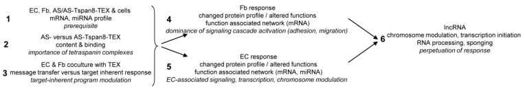
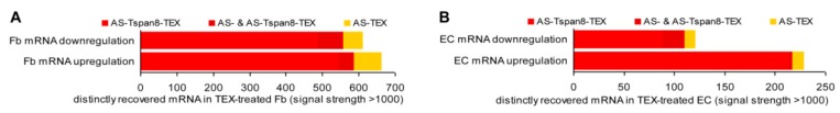
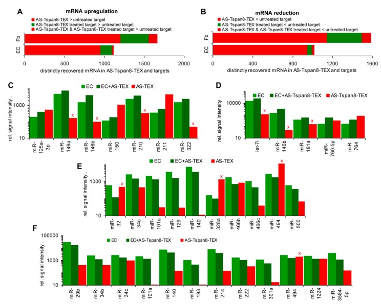
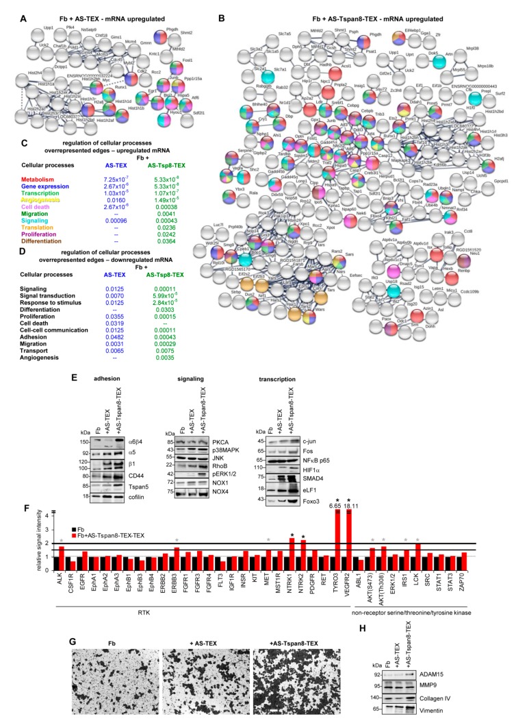
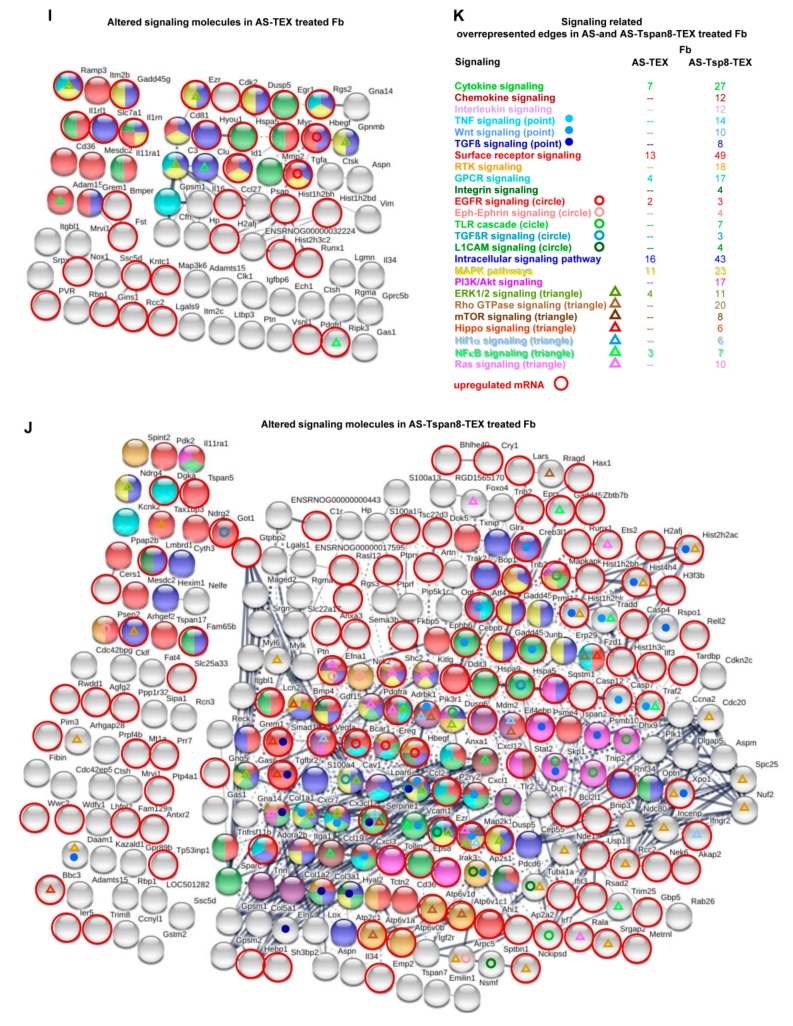
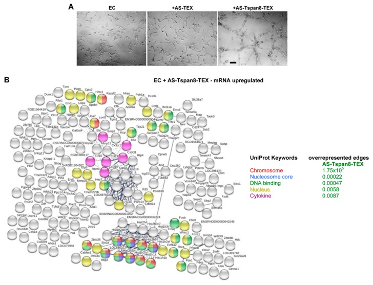
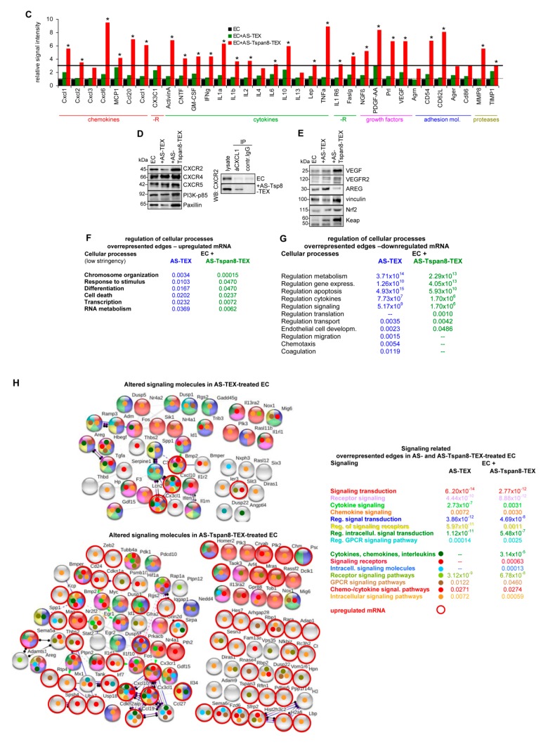
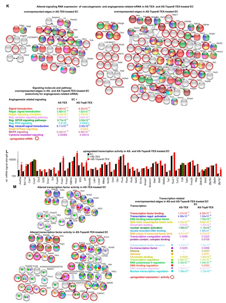
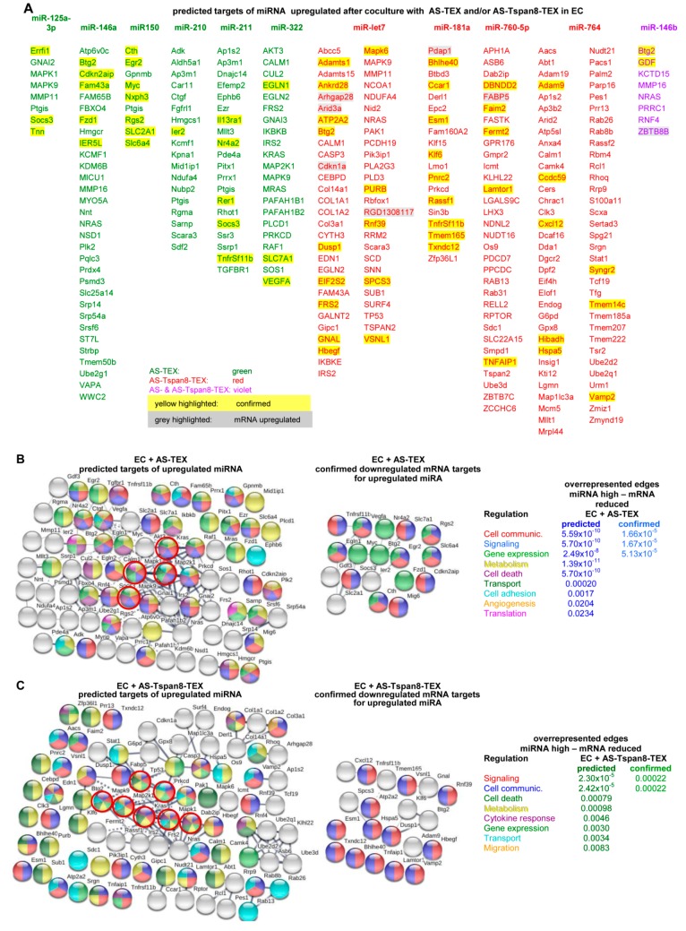
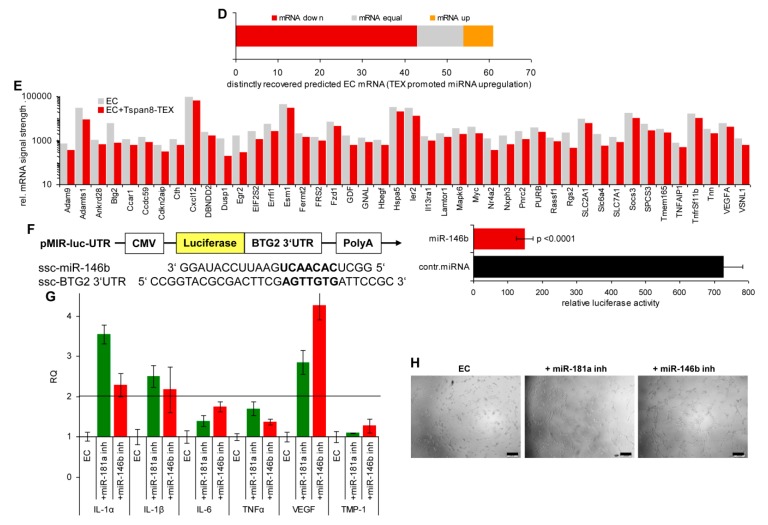
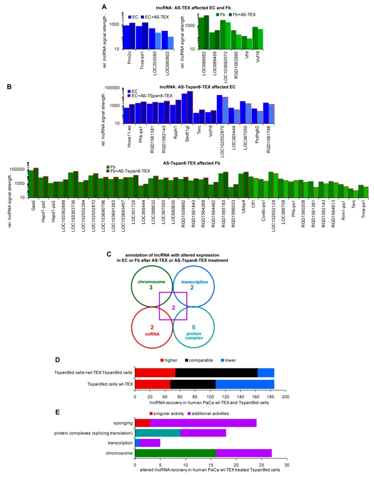
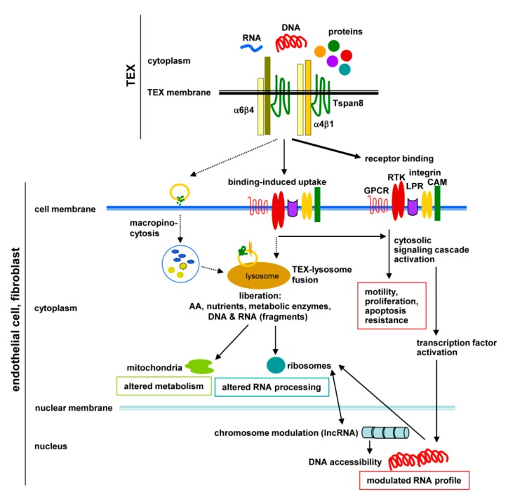
Tumor cell-derived extracellular vesicles (TEX) expressing tetraspanin Tspan8-alpha4/beta1 support angiogenesis. Tspan8-alpha6/beta4 facilitates lung premetastatic niche establishment. TEX-promoted target reprogramming is still being disputed, we explored rat endothelial cell (EC) and lung fibroblast (Fb) mRNA and miRNA profile changes after coculture with TEX. TEX were derived from non-metastatic BSp73AS (AS) or metastatic BSp73ASML (ASML) rat tumor lines transfected with Tspan8 (AS-Tspan8) or Tspan8-shRNA (ASML-Tspan8kd). mRNA was analyzed by deep sequencing and miRNA by array analysis of EC and Fb before and after coculture with TEX. EC and Fb responded more vigorously to AS-Tspan8- than AS-TEX. Though EC and Fb responses differed, both cell lines predominantly responded to membrane receptor activation with upregulation and activation of signaling molecules and transcription factors. Minor TEX-initiated changes in the miRNA profile relied, at least partly, on long noncoding RNA (lncRNA) that also affected chromosome organization and mRNA processing. These analyses uncovered three important points. TEX activate target cell autonomous programs. Responses are initiated by TEX targeting units and are target cell-specific. The strong TEX-promoted lncRNA impact reflects lncRNA shuttling and location-dependent distinct activities. These informations urge for an in depth exploration on the mode of TEX-initiated target cell-specific remodeling including, as a major factor, lncRNA.

Keywords: endothelial cells; fibroblasts; mRNA; message transfer; ncRNA; non-transformed target remodeling; tetraspanin 8; tumor exosomes.


Conflict of interest statement

The authors declare no conflict of interest.

Figures

Figure 1
Figure 1
Experimental workflow.
Figure 2
Figure 2
Distinctly recovered mRNA in fibroblasts and endothelial cells after coculture with tumor cell-derived small extracellular vesicles (TEX). Endothelial cells (EC) and fibroblasts (Fb) were cocultured for 2 d–3 d with 30 µg/mL BSp73AS (AS)- or AS-Tspan8-TEX. mRNA were isolated and subjected to deep sequencing (DS). (A) Fb mRNA and (B) EC mRNA ≥2-fold up- or downregulated by coculture with AS-, AS-Tspan8- or both AS- and AS-Tspan8-TEX. Fb mRNA responds significantly more frequently to coculture with TEX than EC mRNA. Both respond more abundantly to AS-Tspan8- than AS-TEX. AS- and AS-Tspan8-TEX not differing significantly points towards TEX stimulating target cell autonomous programs.
Figure 3
Figure 3
mRNA and miRNA in TEX, TEX-treated fibroblasts, and endothelial cells in comparison to untreated cells. (A) higher mRNA recovery in AS-Tspan8-TEX, AS-Tspan8-TEX-treated Fb or EC, or both TEX and TEX-treated Fb or EC than in untreated target cells (mRNA signal strength of ≥1000, ≥2-fold differences). (B) Corresponding analyses to (A) for reduced mRNA recovery. (C,D) miRNA upregulated in AS- or AS-Tspan8-TEX-treated EC; (E,F) miRNA downregulated in AS- or AS-Tspan8-TEX-treated EC (signal strength ≥500, ≥2-fold change), (CF) Expression in TEX is included for comparison. miRNA expression opposing the recovery in TEX-treated EC is indicated by a red x.
Figure 4
Figure 4
Signaling pathway integration of mRNA up- or downregulated in TEX-treated fibroblasts. mRNA was evaluated in AS- and AS-Tspan8-TEX-treated Fb for up- or downregulation compared to untreated Fb. (AD) STRING functional protein analysis included mRNA with a signal strength ≥1000 and ≥2-fold difference between untreated and TEX-treated Fb, only connected nodes being shown (A) mRNA upregulated in AS-TEX- and (B) AS-Tspan8-TEX-treated Fb; (C) engagement of overrepresented edges of upregulated mRNA in the regulation of biological processes; (D) engagement of overrepresented edges of downregulated mRNA (see Figure S5D) in the regulation of biological processes. (E) WB of indicated proteins in untreated, AS- and AS-Tspan8-TEX-treated Fb; (F) RTK and non-RTK protein array of Fb and AS-Tspan8-TEX-treated Fb. The mean of duplicates is shown, signal intensity being adjusted to 1 for untreated Fb. A significant increase in signal strength ≥1.5-fold is indicated by a grey * and by ≥2-fold by a black *; (G) Fb were seeded on top of matrigel-coated transwell plates that contained RPMI/10% FCS or RPMI/30µg/mL AS- or AS-Tspan8-TEX. After 48 h, cells at the lower membrane site were fixed and stained with crystal violet, light microscopy appearance (scale bar: 20 µm). (H) WB of the indicated proteins in Fb, AS- and AS-Tspan8-TEX-treated Fb; (IK) STRING functional protein analysis for signaling engaged molecules up- or downregulated in (I) AS- and (J) AS-Tspan8-TEX-treated Fb, upregulated mRNA being indicated by a red circle; (K) overrepresented edges for signaling-related molecules and processes, the number of mRNA in overrepresented edges is shown. Full name of gene symbols: Table S10.
Figure 4
Figure 4
Signaling pathway integration of mRNA up- or downregulated in TEX-treated fibroblasts. mRNA was evaluated in AS- and AS-Tspan8-TEX-treated Fb for up- or downregulation compared to untreated Fb. (AD) STRING functional protein analysis included mRNA with a signal strength ≥1000 and ≥2-fold difference between untreated and TEX-treated Fb, only connected nodes being shown (A) mRNA upregulated in AS-TEX- and (B) AS-Tspan8-TEX-treated Fb; (C) engagement of overrepresented edges of upregulated mRNA in the regulation of biological processes; (D) engagement of overrepresented edges of downregulated mRNA (see Figure S5D) in the regulation of biological processes. (E) WB of indicated proteins in untreated, AS- and AS-Tspan8-TEX-treated Fb; (F) RTK and non-RTK protein array of Fb and AS-Tspan8-TEX-treated Fb. The mean of duplicates is shown, signal intensity being adjusted to 1 for untreated Fb. A significant increase in signal strength ≥1.5-fold is indicated by a grey * and by ≥2-fold by a black *; (G) Fb were seeded on top of matrigel-coated transwell plates that contained RPMI/10% FCS or RPMI/30µg/mL AS- or AS-Tspan8-TEX. After 48 h, cells at the lower membrane site were fixed and stained with crystal violet, light microscopy appearance (scale bar: 20 µm). (H) WB of the indicated proteins in Fb, AS- and AS-Tspan8-TEX-treated Fb; (IK) STRING functional protein analysis for signaling engaged molecules up- or downregulated in (I) AS- and (J) AS-Tspan8-TEX-treated Fb, upregulated mRNA being indicated by a red circle; (K) overrepresented edges for signaling-related molecules and processes, the number of mRNA in overrepresented edges is shown. Full name of gene symbols: Table S10.
Figure 5
Figure 5
Angiogenesis-related mRNA modulation and associated signaling activation in TEX-treated endothelial cells. (A) EC were cultured for 48 h in matrigel in the presence of AS- or AS-Tspan8-TEX (30 µg/mL). Growth and spriting was evaluated by light microscopy (scale bar: 20 µm); (B) clusters of mRNA (STRING functional protein analysis) of AS-Tspan8-TEX-treated EC and indication of overrepresented edges according to UniProt Keywords; (C) protein array of chemokines, cytokines, growth factors, receptors, adhesion molecules, and proteases of AS- and AS-Tspan8-TEX-treated EC. Signal strength of duplicates was evaluated by imageJ and is presented adjusting signal strength of EC = 1. The signal strength particularly of cytokines being very low, only >3-fold changes between untreated vs. AS- or AS-Tspan8-TEX-treated EC were considered as significant: *. (D) Western blot (WB) of chemokine receptors, PI3K-p85 and paxillin in untreated and AS- or AS-Tspan8-TEX-treated EC and immunoprecipitation of CXCR2 with CXCL1 in untreated and AS-Tspan8-TEX-treated EC; lysate control and immunoprecipitation with control IgG are included; (E) WB of proteins playing a central role in cytokine/growth factor activation and transcription of related genes. (F,G) Overrepresented edges according to biological processes in AS- and AS-Tspan8-TEX-treated EC, STRING functional protein analysis being performed under less stringent conditions (signal strength ≥500, fold increase: ≥1.5-fold); (F) mRNA upregulated and (G) mRNA downregulated after AS- or AS-Tspan8-TEX treatment. (H) Signaling engaged molecules were selected by Panther Gene list. Altered signaling molecule and signaling regulating mRNA in AS- and AS-Tspan8-TEX treated EC including the incidence of overrepresented edges was evaluated by String functional protein analysis. Upregulated mRNA is indicated by a red circle. (I) Recovery of vasculo-/angiogenesis-related mRNA in AS- and AS-Tspan8-TEX-treated EC, genes affected by AS- and AS-Tspan8-TEX: underlined; inhibitory genes: white bars, mRNA interfering with smooth muscle development: SM. (J) Altered vasculo-/angiogenesis related mRNA were analyzed by STRING for engagement into selective vasculo-/angiogenesis-related functions; upregulated mRNA is marked by a red circle. (K) Altered signaling-related RNA recovery selectively for vasculo-/angiogenesis related mRNA was evaluated according to (J). (L) Transcription factor and transcription initiating molecule mRNA recovery in AS- and AS-Tspan8-TEX-treated EC; >2-fold differences: filled bars, >3-fold differences: red *. (M) Transcription-related molecule overrepresentation as evaluated by STRING; upregulated mRNA is marked by a red circle. Full name of gene symbols: Table S10.
Figure 5
Figure 5
Angiogenesis-related mRNA modulation and associated signaling activation in TEX-treated endothelial cells. (A) EC were cultured for 48 h in matrigel in the presence of AS- or AS-Tspan8-TEX (30 µg/mL). Growth and spriting was evaluated by light microscopy (scale bar: 20 µm); (B) clusters of mRNA (STRING functional protein analysis) of AS-Tspan8-TEX-treated EC and indication of overrepresented edges according to UniProt Keywords; (C) protein array of chemokines, cytokines, growth factors, receptors, adhesion molecules, and proteases of AS- and AS-Tspan8-TEX-treated EC. Signal strength of duplicates was evaluated by imageJ and is presented adjusting signal strength of EC = 1. The signal strength particularly of cytokines being very low, only >3-fold changes between untreated vs. AS- or AS-Tspan8-TEX-treated EC were considered as significant: *. (D) Western blot (WB) of chemokine receptors, PI3K-p85 and paxillin in untreated and AS- or AS-Tspan8-TEX-treated EC and immunoprecipitation of CXCR2 with CXCL1 in untreated and AS-Tspan8-TEX-treated EC; lysate control and immunoprecipitation with control IgG are included; (E) WB of proteins playing a central role in cytokine/growth factor activation and transcription of related genes. (F,G) Overrepresented edges according to biological processes in AS- and AS-Tspan8-TEX-treated EC, STRING functional protein analysis being performed under less stringent conditions (signal strength ≥500, fold increase: ≥1.5-fold); (F) mRNA upregulated and (G) mRNA downregulated after AS- or AS-Tspan8-TEX treatment. (H) Signaling engaged molecules were selected by Panther Gene list. Altered signaling molecule and signaling regulating mRNA in AS- and AS-Tspan8-TEX treated EC including the incidence of overrepresented edges was evaluated by String functional protein analysis. Upregulated mRNA is indicated by a red circle. (I) Recovery of vasculo-/angiogenesis-related mRNA in AS- and AS-Tspan8-TEX-treated EC, genes affected by AS- and AS-Tspan8-TEX: underlined; inhibitory genes: white bars, mRNA interfering with smooth muscle development: SM. (J) Altered vasculo-/angiogenesis related mRNA were analyzed by STRING for engagement into selective vasculo-/angiogenesis-related functions; upregulated mRNA is marked by a red circle. (K) Altered signaling-related RNA recovery selectively for vasculo-/angiogenesis related mRNA was evaluated according to (J). (L) Transcription factor and transcription initiating molecule mRNA recovery in AS- and AS-Tspan8-TEX-treated EC; >2-fold differences: filled bars, >3-fold differences: red *. (M) Transcription-related molecule overrepresentation as evaluated by STRING; upregulated mRNA is marked by a red circle. Full name of gene symbols: Table S10.
Figure 6
Figure 6
Connectivity of target mRNA of upregulated miRNA in TEX-treated endothelial cells. Predicted targets of miRNA upregulated after AS- or AS-Tspan8-TEX treatment were searched for by http://www.microrna.org and http://www.targetscan.org. (A) Predicted mRNA of miRNA expressed at a higher level in EC after coculture with TEX. The threshold level of mRNA was set to a signal strength of ≥500 and a ≥1.5-fold decrease; miRNA upregulated after AS-TEX treatment: green, after AS-Tspan8-TEX treatment: red, after AS- and AS-Tspan8-TEX treatment: violet; predicted mRNA reduction: highlighted yellow, predicted mRNA upregulation: highlighted grey. (B) Spring functional networks of predicted and confirmed downregulated mRNA corresponding to upregulated miRNA after AS-TEX treatment and some significantly overrepresented edges; (C) corresponding analysis to (B) for AS-Tspan8-TEX-promoted upregulated miRNA; (B,C) color of filled circles corresponds to the process of overrepresented edges; circles indicate strongly connected genes; unconnected nodes that were not engaged in the indicated processes were omitted. (D) Summary of mRNA recovery in AS-Tspan8-TEX-treated EC displaying increased miRNA levels and (E) list of downregulated mRNA; (F) Confirmation of BTG2 downregulation by miR-146b as revealed by a double luciferase assay. (G) Confirmation of miR-181a and miR-146b functional activity by culturing EC for 48 h with miR-181a or miR-146b inhibitors or a negative control inhibitor. RNA was isolated and evaluated by qRT-PCR for IL1α, IL1β, IL6, TNFα, VEGF, and TIMP1 expression; relative quantification (RQ) values±SD of triplicates, RQ value of EC cocultured with the negative control inhibitor being set as 1. A 2-fold increase was accepted as significant. (H) EC cultured with miRNA inhibitors as in (G) were seeded on matrigel-precoated plates and incubated with conditioned medium at 37 °C, 5% CO2 in air according to the supplier’s suggestion. Growth and tube formation was evaluated after 8h of culture by light microscopy (scale bar: 200 µm). Full name of gene symbols: Table S10.
Figure 6
Figure 6
Connectivity of target mRNA of upregulated miRNA in TEX-treated endothelial cells. Predicted targets of miRNA upregulated after AS- or AS-Tspan8-TEX treatment were searched for by http://www.microrna.org and http://www.targetscan.org. (A) Predicted mRNA of miRNA expressed at a higher level in EC after coculture with TEX. The threshold level of mRNA was set to a signal strength of ≥500 and a ≥1.5-fold decrease; miRNA upregulated after AS-TEX treatment: green, after AS-Tspan8-TEX treatment: red, after AS- and AS-Tspan8-TEX treatment: violet; predicted mRNA reduction: highlighted yellow, predicted mRNA upregulation: highlighted grey. (B) Spring functional networks of predicted and confirmed downregulated mRNA corresponding to upregulated miRNA after AS-TEX treatment and some significantly overrepresented edges; (C) corresponding analysis to (B) for AS-Tspan8-TEX-promoted upregulated miRNA; (B,C) color of filled circles corresponds to the process of overrepresented edges; circles indicate strongly connected genes; unconnected nodes that were not engaged in the indicated processes were omitted. (D) Summary of mRNA recovery in AS-Tspan8-TEX-treated EC displaying increased miRNA levels and (E) list of downregulated mRNA; (F) Confirmation of BTG2 downregulation by miR-146b as revealed by a double luciferase assay. (G) Confirmation of miR-181a and miR-146b functional activity by culturing EC for 48 h with miR-181a or miR-146b inhibitors or a negative control inhibitor. RNA was isolated and evaluated by qRT-PCR for IL1α, IL1β, IL6, TNFα, VEGF, and TIMP1 expression; relative quantification (RQ) values±SD of triplicates, RQ value of EC cocultured with the negative control inhibitor being set as 1. A 2-fold increase was accepted as significant. (H) EC cultured with miRNA inhibitors as in (G) were seeded on matrigel-precoated plates and incubated with conditioned medium at 37 °C, 5% CO2 in air according to the supplier’s suggestion. Growth and tube formation was evaluated after 8h of culture by light microscopy (scale bar: 200 µm). Full name of gene symbols: Table S10.
Figure 7
Figure 7
Impact of TEX-promoted long noncoding RNA (lncRNA) up- or downregulation on fibroblasts and endothelial cells. Different lncRNA recovery in EC and Fb after coculture with (A) AS-TEX or (B) AS-Tspan8-TEX. (C) Functional assignment of 14/52 lncRNA that expression was altered in EC or Fb after cocultures with TEX. (D) Comparison of lncRNA recovery in human pancreatic adenocarcinoma (PaCa) Tspan8kd cells to wild type (wt)-TEX and of wt-TEX-treated Tspan8kd cells to Tspan8kd cells. (E) Functional assignment of human lncRNA displaying altered expression in wt-TEX-treated Tspan8kd cells. Full name of gene symbols: Tables S8 and S9G.
Figure 8
Figure 8
Coordinated view of Tspan8-TEX-initiated non-transformed cell remodeling. Tspan8-TEX maintain the tetraspanin-associated protein clusters, which facilitate receptor ligand binding that promotes receptor and downstream signaling cascade activation. Alternatively, receptor clustering and activation induces internalization with rarely observed for tetraspanin, subsequent exosome digestion in lysosomes and reutilization of the released breakdown product. Persistence of Tspan8-TEX promoted target cell modulation relies predominantly on activated transcription factor activity modulating the RNA profile. TEX activity is strongly supported by lncRNA that affects chromosomes/DNA accessibility and by forward-backward shuttling between nucleus, ribosomes, and cytoplasm creates circles maintaining remodeling.

Similar articles

Cited by

References

    1. Yáñez-Mó M., Siljander P.R., Andreu Z., Zavec A.B., Borràs F.E., Buzas E.I., Buzas K., Casal E., Cappello F., Carvalho J., et al. Biological properties of extracellular vesicles and their physiological functions. J. Extracell. Vesicles. 2015;4:27066. doi: 10.3402/jev.v4.27066. - DOI - PMC - PubMed
    1. Lindoso R.S., Sandim V., Collino F., Carvalho A.B., Dias J., da Costa M.R., Zingali R.B., Vieyra A. Proteomics of cell-cell interactions in health and disease. Proteomics. 2016;16:328–344. doi: 10.1002/pmic.201500341. - DOI - PubMed
    1. Shang M., Ji J.S., Song C., Gao B.J., Jin J.G., Kuo W.P., Kang H. Extracellular Vesicles: A Brief Overview and Its Role in Precision Medicine. Methods Mol. Biol. 2017;1660:1–14. doi: 10.1007/978-1-4939-7253-1_1. - DOI - PubMed
    1. Gulei D., Irimie A.I., Cojocneanu-Petric R., Schultze J.L., Berindan-Neagoe I. Exosomes-Small Players, Big Sound. Bioconjug. Chem. 2018;29:635–648. doi: 10.1021/acs.bioconjchem.8b00003. - DOI - PubMed
    1. Wang Z., Zöller M. Exosomes, metastases, and the miracle of cancer stem cell markers. Cancer Metastasis Rev. 2019;38:259–295. doi: 10.1007/s10555-019-09793-6. - DOI - PubMed

Publication types

MeSH terms