. 2020 Jan 29;9(2):86.
doi: 10.3390/pathogens9020086.

Increasing the Safety Profile of the Master Donor Live Attenuated Influenza Vaccine

Affiliations

Increasing the Safety Profile of the Master Donor Live Attenuated Influenza Vaccine

Thomas A Hilimire et al. Pathogens. .

Abstract

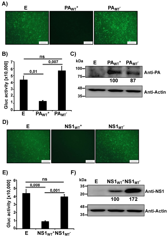
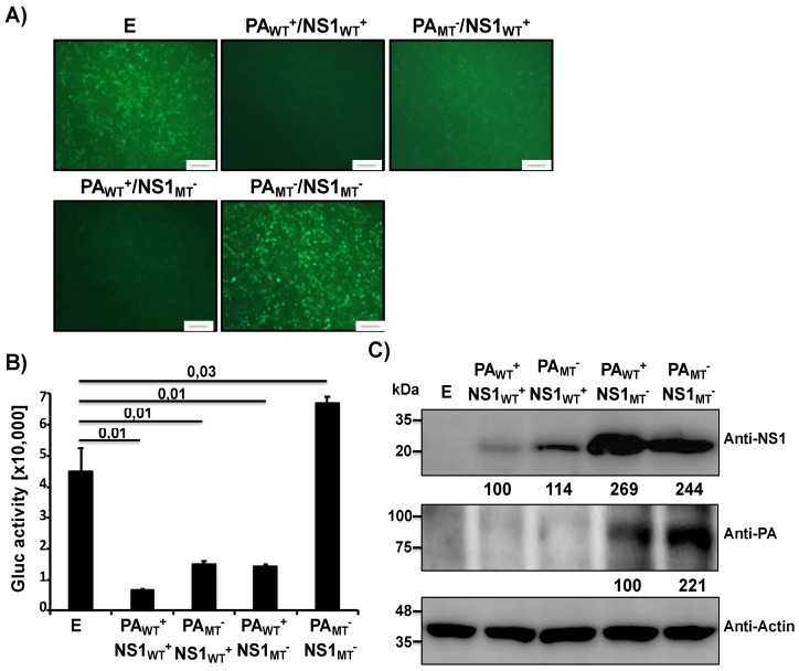
Seasonal influenza epidemics remain one of the largest public health burdens nowadays. The best and most effective strategy to date in preventing influenza infection is a worldwide vaccination campaign. Currently, two vaccines are available to the public for the treatment of influenza infection, the chemically Inactivated Influenza Vaccine (IIV) and the Live Attenuated Influenza Vaccine (LAIV). However, the LAIV is not recommended for parts of the population, such as children under the age of two, immunocompromised individuals, the elderly, and pregnant adults. In order to improve the safety of the LAIV and make it available to more of the population, we sought to further attenuate the LAIV. In this study, we demonstrate that the influenza A virus (IAV) master donor virus (MDV) A/Ann Arbor/6/60 H2N2 LAIV can inhibit host gene expression using both the PA-X and NS1 proteins. Furthermore, we show that by removing PA-X, we can limit the replication of the MDV LAIV in a mouse model, while maintaining full protective efficacy. This work demonstrates a broadly applicable strategy of tuning the amount of host antiviral responses induced by the IAV MDV for the development of newer and safer LAIVs. Moreover, our results also demonstrate, for the first time, the feasibility of genetically manipulating the backbone of the IAV MDV to improve the efficacy of the current IAV LAIV.

Keywords: immunogenicity; influenza; live attenuated influenza vaccine; master donor virus; protection efficacy; vaccine safety; virus–host interaction.


Conflict of interest statement

The authors declare no conflict of interest.

Figures

Figure 1
Figure 1
Ability of master donor virus (MDV) A/AA/6/60 live attenuated influenza vaccine (LAIV) PA and NS1 proteins to block host gene expression: Human 293T cells (5 × 10⁵, 12-well plates, triplicates) were transiently co-transfected, using LPF2000, with expression plasmids encoding GFP and Gluc under the control of a chicken beta actin promoter (pCAGGS GFP and pCAGGS Gluc, respectively) together with pDZ plasmids encoding wild-type (WT+) or mutant (MT) PA or NS1 proteins; or empty (E) plasmid as control. At 24 h post-transfection (p.t.), cells were analyzed by GFP expression (A,D) under a fluorescent microscope and by Gluc activity (B,E) from tissue culture supernatants (TCS) using a luminometer. Representative images are shown. Scale bar = 100 μm. Results represent the means and SDs of triplicate values. Protein expression from cell lysates was evaluated by Western blot (C,F) using specific antibodies for PA (C) or NS1 (F), or actin as the loading control. Molecular markers are indicated on the left. Western blots were quantified by densitometry using the software ImageJ. Relative band intensities (as described in Materials and Methods) are indicated. ns, not statistical.
Figure 2
Figure 2
Ability of MDV A/AA/6/60 LAIV PA-X and NS1 proteins to block host gene expression in combination: Human 293T cells (5 × 10⁵, 12-well plates, triplicates) were transiently co-transfected, using LPF2000, with pCAGGS GFP and pCAGGS Gluc plasmids together with the indicated combination of the pDZ plasmids encoding WT+ or MT PA and NS1 proteins; or empty (E) plasmid as control. At 24 h p.t., cells were analyzed for GFP expression under a fluorescent microscope (A) and for Gluc activity in the TCS using a luminometer (B). Representative images are shown. Scale bar = 100 μm. Results represent the means and SDs of triplicate values. Protein expression from cell lysates was evaluated by Western blot using specific antibodies for PA and NS1 proteins (C). Actin was used as the loading control. Molecular markers are noted on the left. Western blots were quantified by densitometry using the software ImageJ. Relative band intensities (as described in Materials and Methods) are indicated.
Figure 3
Figure 3
Schematic representation of the recombinant MDV A/AA/6/60 LAIVs. (A) Schematic representation of the MDV A/AA/6/60 WT and mutant LAIVs and their gene constellations: Recombinant A/AA/6/60 LAIVs with WT+ and MT segments are indicated with gray or black lines, respectively. PAWT+/NS1WT+: virus containing WT PA (UCC UUU CAU) and NS1 (F103/M106) proteins. PAWT+/NS1MT: virus containing WT PA and mutant NS1 (S103/I106) proteins. PAMT/NS1WT+: virus containing mutant PA (AGC UUC CAC) and WT NS1 proteins. PAMT−/NS1MT+: virus containing both PA and NS1 (mutant proteins. WT+ indicates viral proteins (PA and/or NS1) with the ability to inhibit host gene expression. MT indicates viral proteins (PA or NS1) unable to inhibit host gene expression. (B) Schematic representation of PB2, PB1, NP, PA, and NS1 viral proteins: A/AA/6/60 PB2, PB1, NP, and NS1 proteins (left) with the residues mutated to generate the MDV A/AA/6/60 LAIVs indicated. The WT and MT PA or PA-X viral proteins (right) and the mutations introduced into the frameshift motif (PAMT) to abolish PA-X expression are shown. Numbers on the right indicate the amino acid (aa) length of the viral proteins.
Figure 4
Figure 4
Multicycle growth kinetics and plaque assay of WT and mutant MDV A/AA/6/60 LAIVs. (A) Viral growth kinetics of WT A/AA/6/60 and MDV A/AA/6/60 LAIV at different temperatures: TCS from MDCK cells (5 × 10⁵, 12-well plates, triplicates) infected at low multiplicity of infection (MOI, 0.001) with WT A/AA/6/60 or MDV A/AA/6/60 LAIV at 33 °C, 37 °C, and 39 °C were analyzed at the indicated h p.i. (24, 48, 72, and 96) by immunofocus assay using an anti-NP mAb (HB-65). Data represent the means and SDs of the results determined from triplicate wells. The dashed line indicates the limit of detection (200 fluorescent forming units, FFU/mL). (B,C) Viral growth kinetics of WT MDV A/AA/6/60 LAIV (PAWT+/NS1WT+) and mutant MDV A/AA/6/60 LAIVs (PAWT+/NS1MT, PAMT/NS1WT+, and PAMT/NS1MT): MDCK (B) and A549 (C) cells (5 × 10⁵, 12-well plates, triplicates) were infected (MOI of 0.001 and 0.025, respectively) with the indicated viruses and incubated at 33 °C. TCS were collected at the indicated h p.i. and viral titers were determined by immunofocus assay. Data represent the means and SDs of the results determined from triplicate wells. Dashed lines indicate the limit of detection (200 FFU/mL). *, p < 0.05 using Student’s t test from Microsoft Excel. (D) Plaque assay: MDCK cells (1 × 10⁶, 6-well plates) were infected with the indicated MDV A/AA/60 LAIVs and incubated at 33 °C for 3 days. Plaque phenotypes were visualized by immunostaining using the anti-NP mAb HB-65.
Figure 5
Figure 5
Safety profile of the WT MDV A/AA/6/60 LAIV and mutant MDV A/AA/6/60 LAIVs in vivo: Four-to-six-week-old female C57BL/6 mice (n = 11) were infected i.n. with 1 × 10⁵ FFU of the WT MDV A/AA/6/60 LAIV (PAWT+/NS1WT+) or the mutant MDV A/AA/6/60 LAIVs (PAWT+/NS1MT, PAMT/NS1WT+, and PAMT/NS1MT). Body weight (A) and survival (B) were monitored for 14 days (n = 5). At Days 2 and 4 p.i., C57BL/6-infected mice were sacrificed (n = 3 per time point) and viral titers were determined in lungs (C) and nasal turbinates (D) by immunofocus assay using the anti-NP mAb HB-65. Symbols represent data from individual mice. Bars represent the mean for each group and indicates that the virus was only detected in one or two of the infected mice. N.D. indicates that virus was not detected. Dotted lines represent the limit of detection of the assay (200 FFU/mL).
Figure 6
Figure 6
Humoral responses induced by the MDV A/AA/6/60 LAIVs: Four-to-six-week-old female C57BL/6 mice (n = 5) were infected i.n. with 1 × 10⁵ FFU with the WT MDV A/AA/6/60 LAIV (PAWT+/NS1WT+) or the mutant MDV A/AA/6/60 LAIVs (PAWT+/NS1MT, PAMT/NS1WT+, and PAMT/NS1MT) as indicated. At 21 days p.i., mice were bled, and sera were collected and evaluated for the presences of antibodies against recombinant pH1N1 HA (A) or total viral proteins using cell extracts from pH1N1 virus-infected MDCK cells (B) by ELISA. OD, optical density. (C) HAI titers were calculated from mouse serum. *, p < 0.05 (PAWT+/NS1WT+ vs. the other viruses) using a Student’s t test from Microsoft Excel.
Figure 7
Figure 7
Protective efficacy of the MDV A/AA/6/60 LAIVs: Four-to-six-week-old female C57BL/6 mice (n = 11) were infected i.n. with 1 × 10⁵ FFU of the WT MDV A/AA/6/60 LAIV (PAWT+/NS1WT+) or mutant MDV A/AA/6/60 LAIVs (PAWT+/NS1MT, PAMT/NS1WT+, PAMT/NS1MT), or PBS. At 21 days post-vaccination mice were challenged with 1000X MLD50 of pH1N1. Body weight (A) and mortality (B) were monitored for 14 days (n = 5). At Days 2 and 4 post-challenge, mice were sacrificed (n = 3 per time point) and viral titers in the lungs of infected animals were determined by immunofocus assay using the anti-NP mAb HB-65 (C). Symbols represent data from individual mice. Bars represent the geometric mean for each group and the indicated virus was detected only in one or two mice. The dotted line represents the limit of detection (200 FFU/mL).

Similar articles

Cited by

References

    1. Shaw M.L., Palese P. Othomyxovirdae: the viruses and their replication. Lippincott Williams and Wilkins; Philadelphia, PA, USA: 2007.
    1. Baker S.F., Nogales A., Finch C., Tuffy K.M., Domm W., Perez D.R., Topham D.J., Martinez-Sobrido L. Influenza A and B Virus Intertypic Reassortment through Compatible Viral Packaging Signals. J. Virol. 2014;88:10778–10791. doi: 10.1128/JVI.01440-14. - DOI - PMC - PubMed
    1. Iuliano A.D., Roguski K.M., Chang H.H., Muscatello D.J., Palekar R., Tempia S., Cohen C., Gran J.M., Schanzer D., Cowling B.J., et al. Estimates of global seasonal influenza-associated respiratory mortality: A modelling study. Lancet. 2018;391:1285–1300. doi: 10.1016/S0140-6736(17)33293-2. - DOI - PMC - PubMed
    1. Flannery B., Chung J.R., Belongia E.A., McLean H.Q., Gaglani M., Murthy K., Zimmerman R.K., Nowalk M.P., Jackson M.L., Jackson L.A., et al. Interim Estimates of 2017-18 Seasonal Influenza Vaccine Effectiveness—United States, February 2018. MMWR Morb. Mortal. Wkly. Rep. 2018;67:180–185. doi: 10.15585/mmwr.mm6706a2. - DOI - PMC - PubMed
    1. Mameli C., D’Auria E., Erba P., Nannini P., Zuccotti G.V. Influenza vaccine response: Future perspectives. Expert Opin. Biol. Ther. 2018;18:1–5. doi: 10.1080/14712598.2018.1391786. - DOI - PubMed

LinkOut - more resources