

Vascular Wall-Mesenchymal Stem Cells Differentiation on 3D Biodegradable Highly Porous CaSi-DCPD Doped Poly (α-hydroxy) Acids Scaffolds for Bone Regeneration
- PMID: 32013247
- PMCID: PMC7075175
- DOI: 10.3390/nano10020243
Vascular Wall-Mesenchymal Stem Cells Differentiation on 3D Biodegradable Highly Porous CaSi-DCPD Doped Poly (α-hydroxy) Acids Scaffolds for Bone Regeneration
Abstract
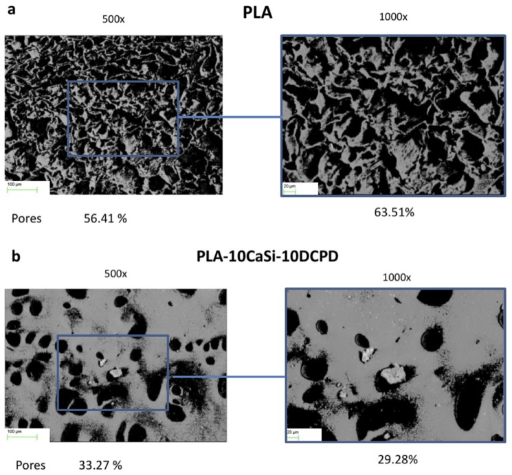
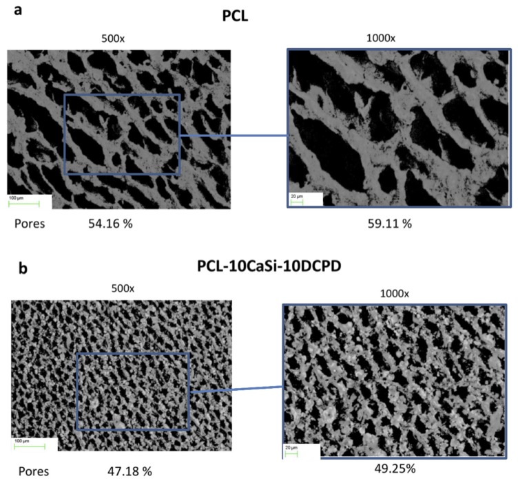
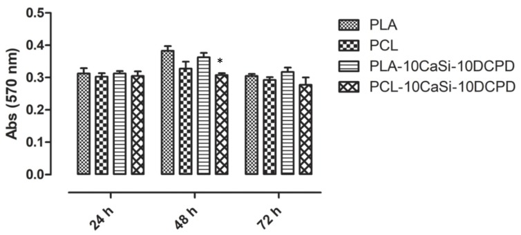
Vascularization is a crucial factor when approaching any engineered tissue. Vascular wall-mesenchymal stem cells are an excellent in vitro model to study vascular remodeling due to their strong angiogenic attitude. This study aimed to demonstrate the angiogenic potential of experimental highly porous scaffolds based on polylactic acid (PLA) or poly-e-caprolactone (PCL) doped with calcium silicates (CaSi) and dicalcium phosphate dihydrate (DCPD), namely PLA-10CaSi-10DCPD and PCL-10CaSi-10DCPD, designed for the regeneration of bone defects. Vascular wall-mesenchymal stem cells (VW-MSCs) derived from pig thoracic aorta were seeded on the scaffolds and the expression of angiogenic markers, i.e. CD90 (mesenchymal stem/stromal cell surface marker), pericyte genes α-SMA (alpha smooth muscle actin), PDGFR-β (platelet-derived growth factor receptor-β), and NG2 (neuron-glial antigen 2) was evaluated. Pure PLA and pure PCL scaffolds and cell culture plastic were used as controls (3D in vitro model vs. 2D in vitro model). The results clearly demonstrated that the vascular wall mesenchymal cells colonized the scaffolds and were metabolically active. Cells, grown in these 3D systems, showed the typical gene expression profile they have in control 2D culture, although with some main quantitative differences. DNA staining and immunofluorescence assay for alpha-tubulin confirmed a cellular presence on both scaffolds. However, VW-MSCs cultured on PLA-10CaSi-10DCPD showed an individual cells growth, whilst on PCL-10CaSi-10DCPD scaffolds VW-MSCs grew in spherical clusters. In conclusion, vascular wall mesenchymal stem cells demonstrated the ability to colonize PLA and PCL scaffolds doped with CaSi-DCPD for new vessels formation and a potential for tissue regeneration.
Keywords: angiogenesis; biobased materials; biodegradable mineral scaffolds; engineered tissue; green biomaterials; green scaffolds; oral bone defects; polylactic acid (PLA), poly-e-caprolactone (PCL); vascular wall–mesenchymal stem cells.
Conflict of interest statement
The authors declare no conflict of interest.
Figures


Similar articles
-
Highly porous polycaprolactone scaffolds doped with calcium silicate and dicalcium phosphate dihydrate designed for bone regeneration.Mater Sci Eng C Mater Biol Appl. 2019 Sep;102:341-361. doi: 10.1016/j.msec.2019.04.040. Epub 2019 Apr 17. Mater Sci Eng C Mater Biol Appl. 2019. PMID: 31147007
-
PLA-Based Mineral-Doped Scaffolds Seeded with Human Periapical Cyst-Derived MSCs: A Promising Tool for Regenerative Healing in Dentistry.Materials (Basel). 2019 Feb 16;12(4):597. doi: 10.3390/ma12040597. Materials (Basel). 2019. PMID: 30781537 Free PMC article.
-
Polylactic acid-based porous scaffolds doped with calcium silicate and dicalcium phosphate dihydrate designed for biomedical application.Mater Sci Eng C Mater Biol Appl. 2018 Jan 1;82:163-181. doi: 10.1016/j.msec.2017.08.040. Epub 2017 Aug 12. Mater Sci Eng C Mater Biol Appl. 2018. PMID: 29025644
-
Effect of poly(3-hydroxyalkanoates) as natural polymers on mesenchymal stem cells.World J Stem Cells. 2019 Oct 26;11(10):764-786. doi: 10.4252/wjsc.v11.i10.764. World J Stem Cells. 2019. PMID: 31692924 Free PMC article. Review.
-
Tissue engineered scaffolds in regenerative medicine.World J Plast Surg. 2014 Jan;3(1):3-7. World J Plast Surg. 2014. PMID: 25489516 Free PMC article. Review.
Cited by
-
PDMS Nano-Modified Scaffolds for Improvement of Stem Cells Proliferation and Differentiation in Microfluidic Platform.Nanomaterials (Basel). 2020 Apr 2;10(4):668. doi: 10.3390/nano10040668. Nanomaterials (Basel). 2020. PMID: 32252384 Free PMC article.
-
Preparation and Characterization of Iron-Doped Tricalcium Silicate-Based Bone Cement as a Bone Repair Material.Materials (Basel). 2020 Aug 19;13(17):3670. doi: 10.3390/ma13173670. Materials (Basel). 2020. PMID: 32825175 Free PMC article.
-
Fabrication Strategies for Bioceramic Scaffolds in Bone Tissue Engineering with Generative Design Applications.Biomimetics (Basel). 2024 Jul 5;9(7):409. doi: 10.3390/biomimetics9070409. Biomimetics (Basel). 2024. PMID: 39056850 Free PMC article. Review.
-
Mineral-Doped Poly(L-lactide) Acid Scaffolds Enriched with Exosomes Improve Osteogenic Commitment of Human Adipose-Derived Mesenchymal Stem Cells.Nanomaterials (Basel). 2020 Feb 29;10(3):432. doi: 10.3390/nano10030432. Nanomaterials (Basel). 2020. PMID: 32121340 Free PMC article.
-
Carbon Nanotubes/Regenerated Silk Composite as a Three-Dimensional Printable Bio-Adhesive Ink with Self-Powering Properties.ACS Appl Mater Interfaces. 2021 May 12;13(18):21007-21017. doi: 10.1021/acsami.1c03288. Epub 2021 May 3. ACS Appl Mater Interfaces. 2021. PMID: 33934601 Free PMC article.
References
LinkOut - more resources
Full Text Sources
Other Literature Sources
Miscellaneous
