Review
. 2020 Jan 29;21(3):868.
doi: 10.3390/ijms21030868.

GM1 Ganglioside Is A Key Factor in Maintaining the Mammalian Neuronal Functions Avoiding Neurodegeneration

Affiliations
Review

GM1 Ganglioside Is A Key Factor in Maintaining the Mammalian Neuronal Functions Avoiding Neurodegeneration

Elena Chiricozzi et al. Int J Mol Sci. .

Abstract

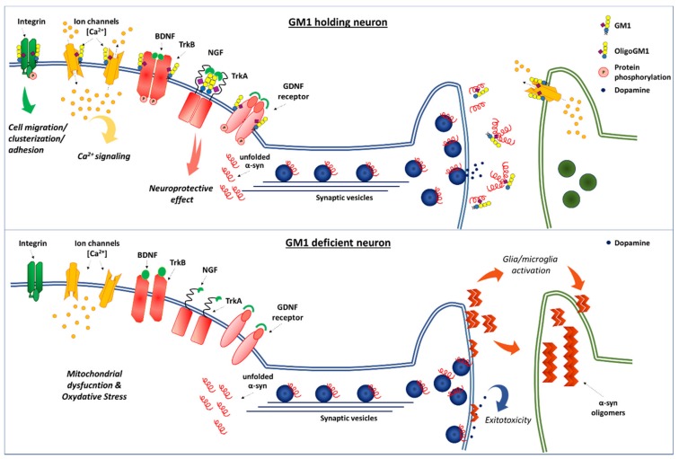
Many species of ganglioside GM1, differing for the sialic acid and ceramide content, have been characterized and their physico-chemical properties have been studied in detail since 1963. Scientists were immediately attracted to the GM1 molecule and have carried on an ever-increasing number of studies to understand its binding properties and its neurotrophic and neuroprotective role. GM1 displays a well balanced amphiphilic behavior that allows to establish strong both hydrophobic and hydrophilic interactions. The peculiar structure of GM1 reduces the fluidity of the plasma membrane which implies a retention and enrichment of the ganglioside in specific membrane domains called lipid rafts. The dynamism of the GM1 oligosaccharide head allows it to assume different conformations and, in this way, to interact through hydrogen or ionic bonds with a wide range of membrane receptors as well as with extracellular ligands. After more than 60 years of studies, it is a milestone that GM1 is one of the main actors in determining the neuronal functions that allows humans to have an intellectual life. The progressive reduction of its biosynthesis along the lifespan is being considered as one of the causes underlying neuronal loss in aged people and severe neuronal decline in neurodegenerative diseases. In this review, we report on the main knowledge on ganglioside GM1, with an emphasis on the recent discoveries about its bioactive component.

Keywords: GM1 ganglioside; GM1 in neurodegeneration; GM1 in neuronal development; GM1 neuronal function; GM1 oligosaccharide function.


Conflict of interest statement

The authors declare no conflict of interest.

Figures

Figure 1
Figure 1
Structure of: N-acetylneuraminic acid, Neu5Ac (A1), N-acetyl-9-acetylneuraminic acid, Neu5,9Ac2 (A2), ganglioside GM1, II3Neu5AcGg4Cer (B), ceramide, Cer R = oligosaccharide chain (C).
Figure 2
Figure 2
Scheme for the preparation of ganglioside GM1 containing [3H] and [14C] isotopically inserted into the lipid and sugar moieties (DDQ, 2,3-dichloro-5,6-dicyanobenzoquinone; PtO2, platinum oxide; NaBH4, sodium borohydride).
Figure 3
Figure 3
Structure of radioactive and photoactivable derivatives of GM1. (A) Structure of [6-3H(IV-Gal)]GM1(Cer-N3); (B) structure of 6-N3(IV-Gal)GM1-[Sph-3-3H]; (C) structure of [6-3H(IV-Gal)]OligoGM1(Glc-N3).
Figure 4
Figure 4
Association of exogenous administered GM1 to cell culture. Ganglioside occurs in water solution in two physical forms, as monomers and micelle. Below a certain concentration, called the critical micelle concentration, gangliosides are found as monomers, whereas above this concentration both micellar and monomer forms are found in equilibrium to each other. The critical micellar concentration for ganglioside GM1 is 10−9 M. GM1 interacts with the cell in 3 different forms, serum labile, trypsin labile and trypsin stabile form [63,64]. (A) Scheme of the association of ganglioside GM1 administered to cells in culture. Micelles interact with the surface and can be removed by serum washing (serum labile form); the form of cell-associated GM1, the trypsin stabile form, corresponds to GM1 single molecules, which is probably present in the plasma membrane external layer and can be removed by trypsin treatment (trypsin labile form of association). Finally monomers are taken up by the cells entering into the cells (trypsin stable form of association). GM1 sugar code is according to Varki et al. [65]. (B) Percent distribution of the three form of GM1 association to rat cerebellar granule cells in culture, as a function of molarity and time [63,64]. As seen in the graph, the proportion of the associated GM1 depends on the incubation time and on the ganglioside concentration of the initial treatment. Decreasing the GM1 concentration, we increase the quantity of GM1 present as monomer instead of GM1 present as micelle in solution and inserted into the cells. Increasing the incubation time, the quantity of GM1 inserted into the cells increases in percentage terms.
Figure 5
Figure 5
Neuronal function dependent by interaction between the GM1 oligosaccharide and proteins [106]. On the top of the image there is a healthy neuron holding with correct GM1 level that sustains neuron homeostasis and also the clearance of α-synuclein. Some of the fundamental neuronal processes that require the association between GM1 oligosaccharide and protein at the plasma membrane level are represented. The viability of dopaminergic neurons depends particularly on the neurotrophic and neuroprotective signaling throughout Trk(s) and RET receptors [2,9]. Fundamental for neuronal survival is also the GM1 collaboration with ion channels (calcium) and integrin receptors [9]. Finally, it is shown also the α-synuclein association with synaptic vesicles and its release in presynaptic region functioning as a regulator of dopamine neurotransmission [197,198,199,200]. Here the correct level of plasma membrane GM1 maintains, in out hypothesis, the α-synuclein in the non-aggregating forms. On the bottom, a neuron with GM1 deficiency and hence a deficiency of its oligosaccharide, that leads to the loss of all the important neurotrophic signals and to α-synuclein aggregation and accumulation. GM1 sugar code is according to Varki et al. [65].

Similar articles

Cited by

References

    1. Kuhn R., Wiegandt H. The Constitution of Gangliosides G-Ii, G-Iii and G-Iv. Z. Naturforsch B. 1963;18:541–543. doi: 10.1515/znb-1963-0708. - DOI - PubMed
    1. Aureli M., Mauri L., Ciampa M.G., Prinetti A., Toffano G., Secchieri C., Sonnino S. GM1 Ganglioside: Past Studies and Future Potential. Mol. Neurobiol. 2016;53:1824–1842. doi: 10.1007/s12035-015-9136-z. - DOI - PubMed
    1. Svennerholm L. The Gangliosides. J. Lipid Res. 1964;5:145–155. - PubMed
    1. Wiegandt H. The structure and the function of gangliosides. Angew Chem. Int. Ed Engl. 1968;7:87–96. doi: 10.1002/anie.196800871. - DOI - PubMed
    1. Holmgren J., Lonnroth I., Mansson J., Svennerholm L. Interaction of cholera toxin and membrane GM1 ganglioside of small intestine. Proc. Natl. Acad. Sci. USA. 1975;72:2520–2524. doi: 10.1073/pnas.72.7.2520. - DOI - PMC - PubMed